• 文献检索
  • 文档翻译
  • 深度研究
  • 学术资讯
  • Suppr Zotero 插件Zotero 插件
  • 邀请有礼
  • 套餐&价格
  • 历史记录
应用&插件
Suppr Zotero 插件Zotero 插件浏览器插件Mac 客户端Windows 客户端微信小程序
定价
高级版会员购买积分包购买API积分包
服务
文献检索文档翻译深度研究API 文档MCP 服务
关于我们
关于 Suppr公司介绍联系我们用户协议隐私条款
关注我们

Suppr 超能文献

核心技术专利:CN118964589B侵权必究
粤ICP备2023148730 号-1Suppr @ 2026

文献检索

告别复杂PubMed语法,用中文像聊天一样搜索,搜遍4000万医学文献。AI智能推荐,让科研检索更轻松。

立即免费搜索

文件翻译

保留排版,准确专业,支持PDF/Word/PPT等文件格式,支持 12+语言互译。

免费翻译文档

深度研究

AI帮你快速写综述,25分钟生成高质量综述,智能提取关键信息,辅助科研写作。

立即免费体验

血管紧张素 II 在重症 COVID-19 中诱导活性氧、DNA 损伤和 T 细胞凋亡。

Angiotensin II induces reactive oxygen species, DNA damage, and T-cell apoptosis in severe COVID-19.

机构信息

Institute of Human Genetics, UMR9002, CNRS, Montpellier University, Montpellier, Nîmes, France.

Immunology Department, Nîmes University Hospital, Nîmes, France.

出版信息

J Allergy Clin Immunol. 2022 Sep;150(3):594-603.e2. doi: 10.1016/j.jaci.2022.06.020. Epub 2022 Jul 14.

DOI:10.1016/j.jaci.2022.06.020
PMID:35841981
原文链接:https://pmc.ncbi.nlm.nih.gov/articles/PMC9278992/
Abstract

BACKGROUND

Lymphopenia is predictive of survival in patients with coronavirus disease 2019 (COVID-19).

OBJECTIVE

The aim of this study was to understand the cause of the lymphocyte count drop in severe forms of severe acute respiratory syndrome coronavirus 2 (SARS-CoV-2) infection.

METHODS

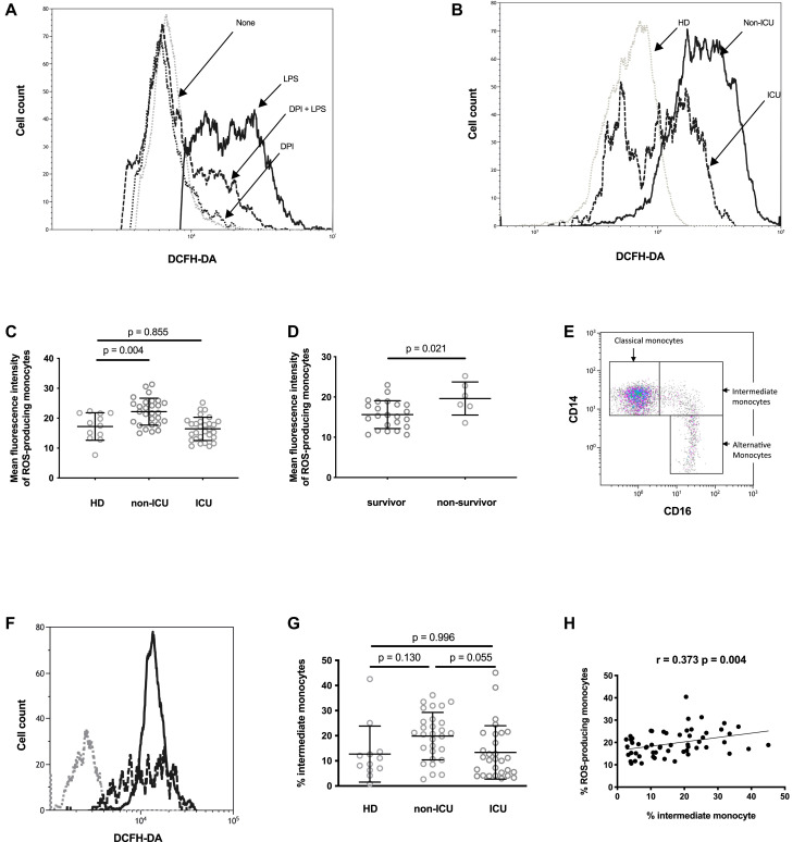
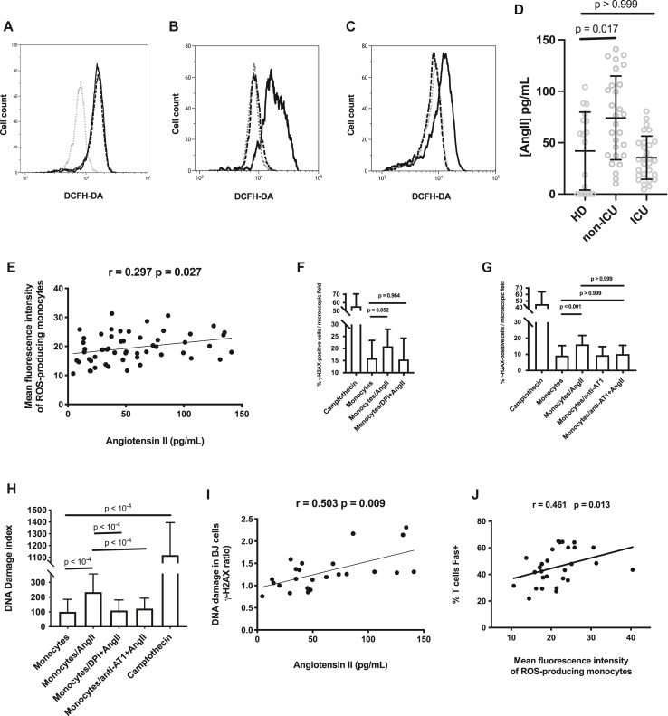
Monocytic production of reactive oxygen species (ROSs) and T-cell apoptosis were measured by flow cytometry, DNA damage in PBMCs was measured by immunofluorescence, and angiotensin II (AngII) was measured by ELISA in patients infected with SARS-CoV-2 at admission to an intensive care unit (ICU) (n = 29) or not admitted to an ICU (n = 29) and in age- and sex-matched healthy controls.

RESULTS

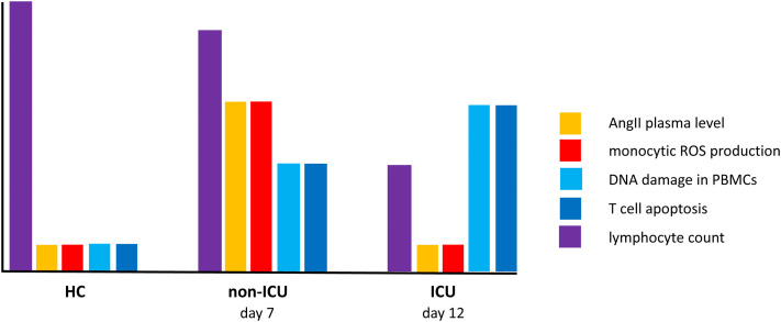
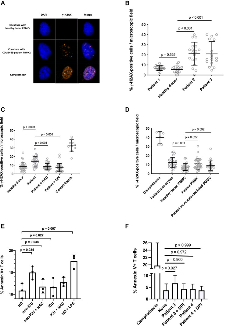
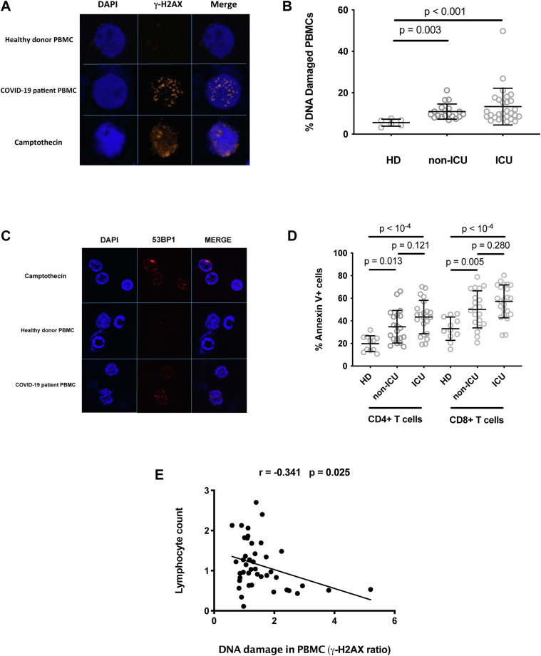
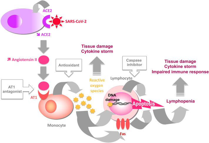
We showed that the monocytes of certain patients with COVID-19 spontaneously released ROSs able to induce DNA damage and apoptosis in neighboring cells. Of note, high ROS production was predictive of death in ICU patients. Accordingly, in most patients, we observed the presence of DNA damage in up to 50% of their PBMCs and T-cell apoptosis. Moreover, the intensity of this DNA damage was linked to lymphopenia. SARS-CoV-2 is known to induce the internalization of its receptor, angiotensin-converting enzyme 2, which is a protease capable of catabolizing AngII. Accordingly, in certain patients with COVID-19 we observed high plasma levels of AngII. When looking for the stimulus responsible for their monocytic ROS production, we revealed that AngII triggers ROS production by monocytes via angiotensin receptor I. ROSs released by AngII-activated monocytes induced DNA damage and apoptosis in neighboring lymphocytes.

CONCLUSION

We conclude that T-cell apoptosis provoked via DNA damage due to the release of monocytic ROSs could play a major role in COVID-19 pathogenesis.

摘要

背景

淋巴细胞减少与 2019 年冠状病毒病(COVID-19)患者的生存有关。

目的

本研究旨在了解严重急性呼吸综合征冠状病毒 2(SARS-CoV-2)感染严重形式中淋巴细胞计数下降的原因。

方法

通过流式细胞术测量单核细胞产生的活性氧(ROS)和 T 细胞凋亡,通过免疫荧光法测量 PBMCs 中的 DNA 损伤,通过 ELISA 测量血管紧张素 II(AngII)在因 SARS-CoV-2 感染而入住重症监护病房(ICU)的患者(n=29)或未入住 ICU 的患者(n=29)和年龄及性别匹配的健康对照者中。

结果

我们表明,某些 COVID-19 患者的单核细胞会自发释放 ROS,能够诱导邻近细胞的 DNA 损伤和凋亡。值得注意的是,高 ROS 产生可预测 ICU 患者的死亡。因此,在大多数患者中,我们观察到多达 50%的 PBMC 和 T 细胞凋亡存在 DNA 损伤。此外,这种 DNA 损伤的强度与淋巴细胞减少有关。众所周知,SARS-CoV-2 会诱导其受体血管紧张素转换酶 2 的内化,血管紧张素转换酶 2 是一种能够分解 AngII 的蛋白酶。因此,在某些 COVID-19 患者中,我们观察到 AngII 的血浆水平较高。当寻找导致其单核细胞 ROS 产生的刺激物时,我们发现 AngII 通过血管紧张素受体 I 触发单核细胞产生 ROS。AngII 激活的单核细胞释放的 ROS 诱导邻近淋巴细胞的 DNA 损伤和凋亡。

结论

我们得出结论,由于单核细胞 ROS 释放引起的 DNA 损伤导致的 T 细胞凋亡可能在 COVID-19 发病机制中起重要作用。

https://cdn.ncbi.nlm.nih.gov/pmc/blobs/0663/9278992/78b9d77b1b12/fx2_lrg.jpg
https://cdn.ncbi.nlm.nih.gov/pmc/blobs/0663/9278992/0efda1b81f35/gr1_lrg.jpg
https://cdn.ncbi.nlm.nih.gov/pmc/blobs/0663/9278992/59363c360a9e/gr2_lrg.jpg
https://cdn.ncbi.nlm.nih.gov/pmc/blobs/0663/9278992/57044e2f7a10/gr3_lrg.jpg
https://cdn.ncbi.nlm.nih.gov/pmc/blobs/0663/9278992/5137e68eab17/gr4_lrg.jpg
https://cdn.ncbi.nlm.nih.gov/pmc/blobs/0663/9278992/cab297f2e9ff/fx1_lrg.jpg
https://cdn.ncbi.nlm.nih.gov/pmc/blobs/0663/9278992/78b9d77b1b12/fx2_lrg.jpg

相似文献

1
Angiotensin II induces reactive oxygen species, DNA damage, and T-cell apoptosis in severe COVID-19.血管紧张素 II 在重症 COVID-19 中诱导活性氧、DNA 损伤和 T 细胞凋亡。
J Allergy Clin Immunol. 2022 Sep;150(3):594-603.e2. doi: 10.1016/j.jaci.2022.06.020. Epub 2022 Jul 14.
2
SARS-CoV-2-associated lymphopenia: possible mechanisms and the role of CD147.SARS-CoV-2 相关的淋巴细胞减少症:可能的机制和 CD147 的作用。
Cell Commun Signal. 2024 Jul 4;22(1):349. doi: 10.1186/s12964-024-01718-3.
3
Angiotensin II Exaggerates SARS-CoV-2 Specific T-Cell Response in Convalescent Individuals following COVID-19.血管紧张素 II 在 COVID-19 康复者中加剧了针对 SARS-CoV-2 的 T 细胞反应。
Int J Mol Sci. 2022 Aug 4;23(15):8669. doi: 10.3390/ijms23158669.
4
Lymphopenia an important immunological abnormality in patients with COVID-19: Possible mechanisms.淋巴细胞减少症是 COVID-19 患者重要的免疫学异常:可能的机制。
Scand J Immunol. 2021 Feb;93(2):e12967. doi: 10.1111/sji.12967. Epub 2020 Sep 14.
5
Systemic redox imbalance in severe COVID-19 patients.严重 COVID-19 患者的全身氧化还原失衡。
Cell Biochem Funct. 2022 Oct;40(7):694-705. doi: 10.1002/cbf.3735. Epub 2022 Aug 18.
6
Linking ACE2 and angiotensin II to pulmonary immunovascular dysregulation in SARS-CoV-2 infection.将 ACE2 和血管紧张素 II 与 SARS-CoV-2 感染中的肺免疫血管失调联系起来。
Int J Infect Dis. 2020 Dec;101:42-45. doi: 10.1016/j.ijid.2020.09.041. Epub 2020 Sep 17.
7
Apoptosis-induced T-cell lymphopenia is related to COVID-19 severity.细胞凋亡诱导的 T 细胞淋巴细胞减少与 COVID-19 严重程度有关。
J Med Virol. 2021 May;93(5):2867-2874. doi: 10.1002/jmv.26742. Epub 2021 Feb 23.
8
Distinct Mitochondria-Mediated T-Cell Apoptosis Responses in Children and Adults With Coronavirus Disease 2019.2019 年冠状病毒病患儿与成人之间存在独特的线粒体介导的 T 细胞凋亡反应。
J Infect Dis. 2021 Oct 28;224(8):1333-1344. doi: 10.1093/infdis/jiab400.
9
T cell apoptosis characterizes severe Covid-19 disease.T 细胞凋亡是严重新冠肺炎疾病的特征。
Cell Death Differ. 2022 Aug;29(8):1486-1499. doi: 10.1038/s41418-022-00936-x. Epub 2022 Jan 22.
10
The clinical and immunological characteristics of COVID-19 patients with delayed SARS-CoV-2 virus clearance.新冠病毒清除延迟患者的临床和免疫学特征。
Immun Inflamm Dis. 2023 Sep;11(9):e999. doi: 10.1002/iid3.999.

引用本文的文献

1
GCRV-II Triggers B and T Lymphocyte Apoptosis via Mitochondrial ROS Pathway.草鱼呼肠孤病毒II型通过线粒体活性氧途径触发B淋巴细胞和T淋巴细胞凋亡。
Viruses. 2025 Jun 30;17(7):930. doi: 10.3390/v17070930.
2
Evaluating the Role of the Renin-angiotensin System in COVID-19: Implications for ACE Inhibitor and ARB Use During SARS-CoV-2 Infection.评估肾素-血管紧张素系统在2019冠状病毒病中的作用:对严重急性呼吸综合征冠状病毒2感染期间使用血管紧张素转换酶抑制剂和血管紧张素Ⅱ受体拮抗剂的启示
J Cell Immunol. 2024;6(6):255-265. doi: 10.33696/immunology.6.213.
3
Thromboembolic Events in the Era of COVID-19: A Detailed Narrative Review.
COVID-19 时代的血栓栓塞事件:详细叙述性综述
Can J Infect Dis Med Microbiol. 2025 Mar 4;2025:3804576. doi: 10.1155/cjid/3804576. eCollection 2025.
4
Mitochondrial Dysfunction and Metabolic Disturbances Induced by Viral Infections.病毒感染诱导的线粒体功能障碍和代谢紊乱。
Cells. 2024 Oct 29;13(21):1789. doi: 10.3390/cells13211789.
5
DNA Damage in Moderate and Severe COVID-19 Cases: Relation to Demographic, Clinical, and Laboratory Parameters.中度和重度 COVID-19 病例中的 DNA 损伤:与人口统计学、临床和实验室参数的关系。
Int J Mol Sci. 2024 Sep 24;25(19):10293. doi: 10.3390/ijms251910293.
6
Melatonin protects against defects induced by methoxychlor in porcine oocyte maturation.褪黑素可防止甲氧滴滴涕诱导的猪卵母细胞成熟缺陷。
Cell Cycle. 2024 Sep 16:1-17. doi: 10.1080/15384101.2024.2402190.
7
SARS-CoV-2-associated lymphopenia: possible mechanisms and the role of CD147.SARS-CoV-2 相关的淋巴细胞减少症:可能的机制和 CD147 的作用。
Cell Commun Signal. 2024 Jul 4;22(1):349. doi: 10.1186/s12964-024-01718-3.
8
T4 apoptosis in the acute phase of SARS-CoV-2 infection predicts long COVID.SARS-CoV-2 感染急性期的 T4 细胞凋亡可预测长新冠。
Front Immunol. 2024 Jan 3;14:1335352. doi: 10.3389/fimmu.2023.1335352. eCollection 2023.
9
Characteristics of hematological parameters on admission in COVID-19 Omicron variant infected in Chinese population: a large-scale retrospective study.奥密克戎变异株感染的中国人群入院时血液学参数特征:一项大规模回顾性研究。
BMC Infect Dis. 2023 Nov 27;23(1):835. doi: 10.1186/s12879-023-08771-2.
10
Myocardial injury and related mortality in hospitalized patients with COVID-19 during the Omicron pandemic: new perspectives and insights.奥密克戎流行期间住院 COVID-19 患者的心肌损伤和相关死亡率:新视角和见解。
Virol Sin. 2023 Dec;38(6):940-950. doi: 10.1016/j.virs.2023.10.005. Epub 2023 Oct 13.