Seo Jeong-Wook, Kim Jung-Sun, Cha Myung-Jin, Yoon Ja Kyoung, Kim Min-Ju, Tsao Hsuan-Ming, Lee Chang-Ha, Oh Seil
Department of Pathology, Incheon Sejong Hospital, Incheon, Korea.
Department of Pathology, Seoul National University, Seoul, Korea.
J Chest Surg. 2022 Oct 5;55(5):364-377. doi: 10.5090/jcs.22.030. Epub 2022 Jul 20.
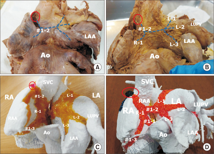
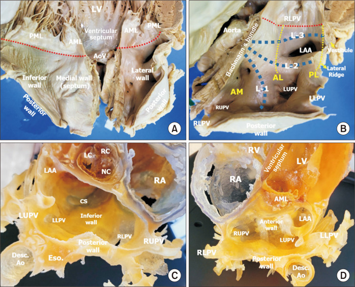
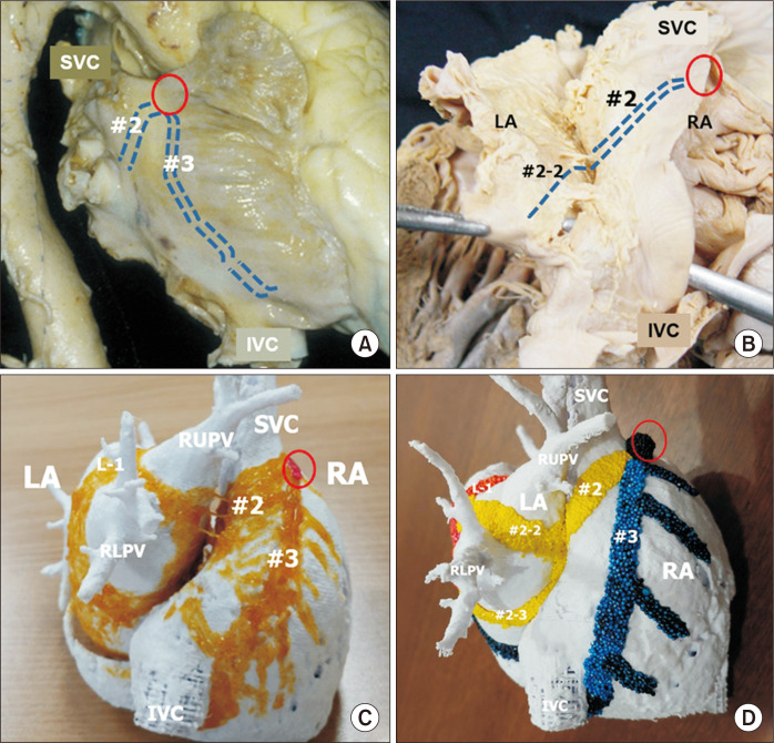
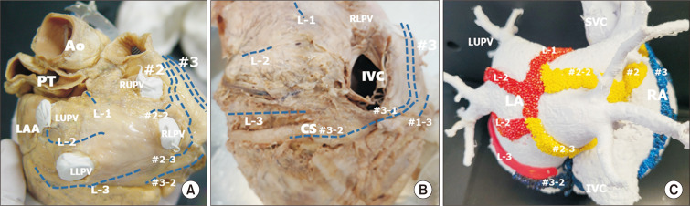
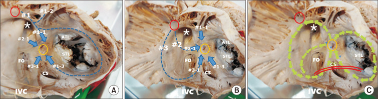
An anatomical understanding of the atrial myocardium is crucial for surgeons and interventionists who treat atrial arrhythmias. We reviewed the anatomy of the inter-nodal and intra-atrial conduction systems. The anterior inter-nodal route (#1) arises from the sinus node and runs through the ventral wall of the atrial chambers. The major branch of route #1 approaches the atrioventricular node from the anterior aspect. Other branches of route #1 are Bachmann's bundle and a vestibular branch around the tricuspid valve. The middle inter-nodal route (#2) begins with a broad span of fibers at the sinus venarum and extends to the superior limbus of the oval fossa. The major branch of route #2 joins with the branch of route #1 at the anterior part of the atrioventricular node. The posterior inter-nodal route (#3) is at the terminal crest and gives rise to many branches at the pectinate muscles of the right atrium and then approaches the posterior atrioventricular node after joining with the vestibular branch of route #1. The branches of the left part of Bachmann's bundle and the branches of the second inter-nodal route form a thin myocardial network at the posterior wall of the left atrium. These anatomical structures could be categorized into major routes and side branches. There are 9 or more anatomical circles in the atrial chambers that could be structural sites for macro re-entry. The implications of normal and abnormal structures of the myocardium for the pathogenesis and treatment of atrial arrhythmias are discussed.
对于治疗房性心律失常的外科医生和介入医生来说,了解心房心肌的解剖结构至关重要。我们回顾了结间和心房内传导系统的解剖结构。前结间径路(#1)起自窦房结,穿过心房腔的前壁。径路#1的主要分支从前侧接近房室结。径路#1的其他分支是巴赫曼束和围绕三尖瓣的前庭支。中间结间径路(#2)始于腔静脉窦处的一大片纤维,延伸至卵圆窝上缘。径路#2的主要分支在房室结前部与径路#1的分支汇合。后结间径路(#3)位于终嵴,在右心房梳状肌处发出许多分支,然后在与径路#1的前庭支汇合后接近后房室结。巴赫曼束左侧分支和第二结间径路分支在左心房后壁形成一个薄的心肌网络。这些解剖结构可分为主要径路和侧支。心房腔内有9个或更多的解剖环路,可能是大折返的结构部位。本文讨论了心肌正常和异常结构对房性心律失常发病机制和治疗的影响。