• 文献检索
  • 文档翻译
  • 深度研究
  • 学术资讯
  • Suppr Zotero 插件Zotero 插件
  • 邀请有礼
  • 套餐&价格
  • 历史记录
应用&插件
Suppr Zotero 插件Zotero 插件浏览器插件Mac 客户端Windows 客户端微信小程序
定价
高级版会员购买积分包购买API积分包
服务
文献检索文档翻译深度研究API 文档MCP 服务
关于我们
关于 Suppr公司介绍联系我们用户协议隐私条款
关注我们

Suppr 超能文献

核心技术专利:CN118964589B侵权必究
粤ICP备2023148730 号-1Suppr @ 2026

文献检索

告别复杂PubMed语法,用中文像聊天一样搜索,搜遍4000万医学文献。AI智能推荐,让科研检索更轻松。

立即免费搜索

文件翻译

保留排版,准确专业,支持PDF/Word/PPT等文件格式,支持 12+语言互译。

免费翻译文档

深度研究

AI帮你快速写综述,25分钟生成高质量综述,智能提取关键信息,辅助科研写作。

立即免费体验

从烧伤增生性瘢痕中分离的真皮微血管内皮细胞的血管生成基因特征和血管通透性。

Angiogenic gene characterization and vessel permeability of dermal microvascular endothelial cells isolated from burn hypertrophic scar.

机构信息

Georgetown University School of Medicine, Washington, DC, USA.

Firefighter's Burn and Surgical Research Laboratory, MedStar Health Research Institute, 110 Irving Street, NW, GHRB, Room 310, Washington, DC, 20010, USA.

出版信息

Sci Rep. 2022 Jul 18;12(1):12222. doi: 10.1038/s41598-022-16376-z.

DOI:10.1038/s41598-022-16376-z
PMID:35851095
原文链接:https://pmc.ncbi.nlm.nih.gov/articles/PMC9293893/
Abstract

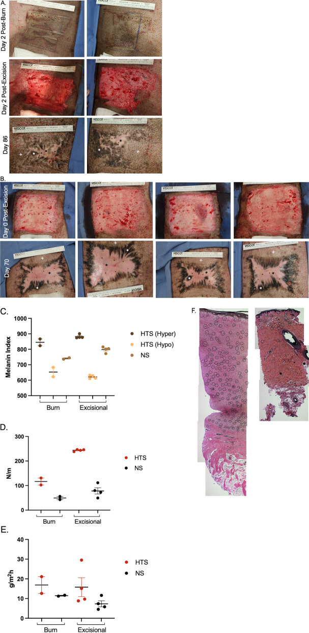
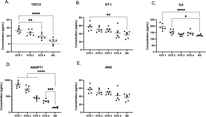
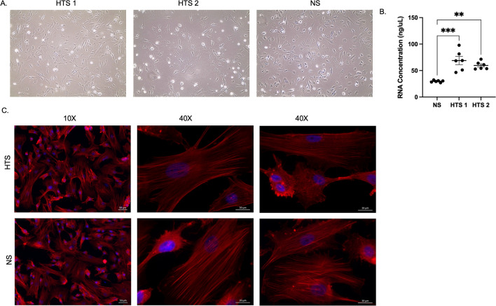
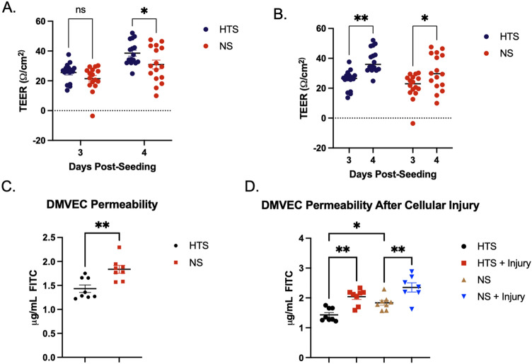
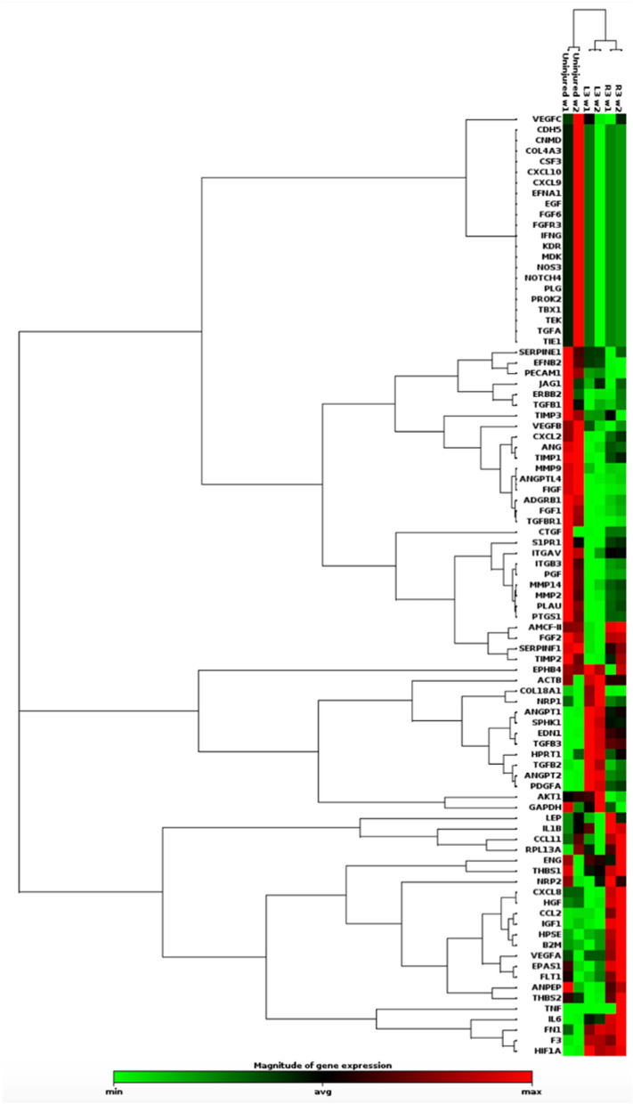
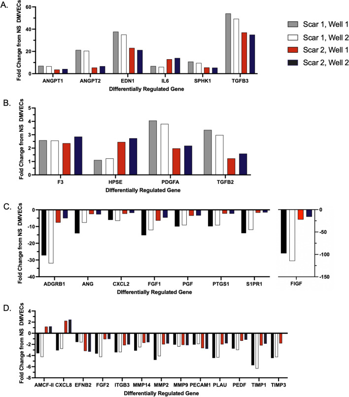
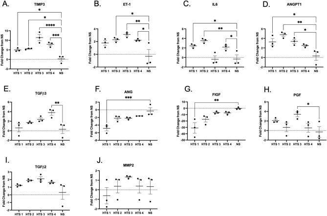
Hypertrophic scar (HTS) formation is a common challenge for patients after burn injury. Dermal microvascular endothelial cells (DMVECs) are an understudied cell type in HTS. An increase in angiogenesis and microvessel density can be observed in HTS. Endothelial dysfunction may play a role in scar development. This study aims to generate a functional and expression profile of HTS DMVECs. We hypothesize that transcript and protein-level responses in HTS DMVECs differ from those in normal skin (NS). HTSs were created in red Duroc pigs. DMVECs were isolated using magnetic-activated cell sorting with ulex europaeus agglutinin 1 (UEA-1) lectin. Separate transwell inserts were used to form monolayers of HTS DMVECs and NS DMVECs. Cell injury was induced and permeability was assessed. Gene expression in HTS DMVECS versus NS DMVECs was measured. Select differentially expressed genes were further investigated. HTS had an increased area density of dermal microvasculature compared to NS. HTS DMVECs were 17.59% less permeable than normal DMVECs (p < 0.05). After injury, NS DMVECs were 28.4% and HTS DMVECs were 18.8% more permeable than uninjured controls (28.4 ± 4.8 vs 18.8 ± 2.8; p = 0.11). PCR array identified 31 differentially expressed genes between HTS and NS DMVECs, of which 10 were upregulated and 21 were downregulated. qRT-PCR and ELISA studies were in accordance with the array. DMVECs expressed a mixed profile of factors that can contribute to and inhibit scar formation. HTS DMVECs have both a discordant response to cellular insults and baseline differences in function, supporting their proposed role in scar pathology. Further investigation of DMVECs is warranted to elucidate their contribution to HTS pathogenesis.

摘要

增生性瘢痕(HTS)的形成是烧伤患者常见的挑战。真皮微血管内皮细胞(DMVECs)是 HTS 中研究较少的细胞类型。在 HTS 中可以观察到血管生成和微血管密度的增加。内皮功能障碍可能在瘢痕形成中起作用。本研究旨在生成 HTS DMVECs 的功能和表达谱。我们假设 HTS DMVECs 的转录和蛋白水平反应与正常皮肤(NS)不同。使用针对 Ulex europaeus agglutinin 1(UEA-1)凝集素的磁激活细胞分选从红色杜洛克猪中分离 DMVECs。使用单独的 Transwell 插入物形成 HTS DMVECs 和 NS DMVECs 的单层。诱导细胞损伤并评估通透性。测量 HTS DMVEC 与 NS DMVEC 中的基因表达。进一步研究了选定的差异表达基因。与 NS 相比,HTS 的真皮微血管面积密度增加。HTS DMVECs 的通透性比正常 DMVECs 低 17.59%(p<0.05)。受伤后,NS DMVECs 的通透性比未受伤对照高 28.4%(28.4±4.8),而 HTS DMVECs 的通透性高 18.8%(18.8±2.8;p=0.11)。PCR 阵列在 HTS 和 NS DMVECs 之间鉴定出 31 个差异表达基因,其中 10 个上调,21 个下调。qRT-PCR 和 ELISA 研究与阵列一致。DMVECs 表达了可促进和抑制瘢痕形成的多种因子的混合谱。HTS DMVECs 对细胞损伤的反应不一致,且功能基线存在差异,支持它们在瘢痕病理学中的预期作用。进一步研究 DMVECs 对于阐明其对 HTS 发病机制的贡献是必要的。

https://cdn.ncbi.nlm.nih.gov/pmc/blobs/d14f/9293893/186f42bbb049/41598_2022_16376_Fig7_HTML.jpg
https://cdn.ncbi.nlm.nih.gov/pmc/blobs/d14f/9293893/8b5340c28adc/41598_2022_16376_Fig1_HTML.jpg
https://cdn.ncbi.nlm.nih.gov/pmc/blobs/d14f/9293893/89ef91243c0d/41598_2022_16376_Fig2_HTML.jpg
https://cdn.ncbi.nlm.nih.gov/pmc/blobs/d14f/9293893/71bb89b0fa2c/41598_2022_16376_Fig3_HTML.jpg
https://cdn.ncbi.nlm.nih.gov/pmc/blobs/d14f/9293893/6cbc8e8beb2f/41598_2022_16376_Fig4_HTML.jpg
https://cdn.ncbi.nlm.nih.gov/pmc/blobs/d14f/9293893/c61047fbcbef/41598_2022_16376_Fig5_HTML.jpg
https://cdn.ncbi.nlm.nih.gov/pmc/blobs/d14f/9293893/78ae84900498/41598_2022_16376_Fig6_HTML.jpg
https://cdn.ncbi.nlm.nih.gov/pmc/blobs/d14f/9293893/186f42bbb049/41598_2022_16376_Fig7_HTML.jpg

相似文献

1
Angiogenic gene characterization and vessel permeability of dermal microvascular endothelial cells isolated from burn hypertrophic scar.从烧伤增生性瘢痕中分离的真皮微血管内皮细胞的血管生成基因特征和血管通透性。
Sci Rep. 2022 Jul 18;12(1):12222. doi: 10.1038/s41598-022-16376-z.
2
Laser Treatment of Hypertrophic Scar in a Porcine Model Induces Change to Epidermal Histoarchitecture That Correlates to Improved Epidermal Barrier Function.激光治疗猪模型肥厚性瘢痕诱导表皮组织形态发生改变,与改善表皮屏障功能相关。
J Burn Care Res. 2023 Jul 5;44(4):758-768. doi: 10.1093/jbcr/irad010.
3
Improved scar in postburn patients following interferon-alpha2b treatment is associated with decreased angiogenesis mediated by vascular endothelial cell growth factor.α-干扰素2b治疗后烧伤患者瘢痕改善与血管内皮细胞生长因子介导的血管生成减少有关。
J Interferon Cytokine Res. 2008 Jul;28(7):423-34. doi: 10.1089/jir.2007.0104.
4
Treatment of hypopigmented burn hypertrophic scars with short-term topical tacrolimus does not lead to repigmentation.短期外用他克莫司治疗色素减退性烧伤增生性瘢痕不会导致色素再沉着。
Lasers Surg Med. 2024 Feb;56(2):175-185. doi: 10.1002/lsm.23754. Epub 2024 Jan 15.
5
Galectin-1 production is elevated in hypertrophic scar.Galectin-1 的产量在增生性瘢痕中升高。
Wound Repair Regen. 2021 Jan;29(1):117-128. doi: 10.1111/wrr.12869. Epub 2020 Oct 26.
6
Stromal cell-derived factor 1 (SDF-1) and its receptor CXCR4 in the formation of postburn hypertrophic scar (HTS).基质细胞衍生因子 1(SDF-1)及其受体 CXCR4 在烧伤后肥厚性瘢痕(HTS)的形成中的作用。
Wound Repair Regen. 2011 Sep-Oct;19(5):568-78. doi: 10.1111/j.1524-475X.2011.00724.x.
7
Autologous adipose-derived regenerative cell therapy modulates development of hypertrophic scarring in a red Duroc porcine model.自体脂肪来源的再生细胞疗法可调节红色杜洛克猪模型中增生性瘢痕的形成。
Stem Cell Res Ther. 2017 Nov 15;8(1):261. doi: 10.1186/s13287-017-0704-1.
8
Toll-like receptors expressed by dermal fibroblasts contribute to hypertrophic scarring.真皮成纤维细胞表达的 Toll 样受体有助于增生性瘢痕形成。
J Cell Physiol. 2011 May;226(5):1265-73. doi: 10.1002/jcp.22454.
9
Endothelial-to-mesenchymal transition contributes to endothelial dysfunction and dermal fibrosis in systemic sclerosis.内皮细胞向间充质转化促进系统性硬皮病的内皮功能障碍和皮肤纤维化。
Ann Rheum Dis. 2017 May;76(5):924-934. doi: 10.1136/annrheumdis-2016-210229. Epub 2017 Jan 6.
10
Comparison of Normal Human Skin and Hypertrophic Scar Tissue Samples of Different Ages, Locations, and Stages of Maturity.不同年龄、部位和成熟阶段的正常人皮肤与肥厚性瘢痕组织样本的比较。
Ann Plast Surg. 2024 Oct 1;93(4):434-442. doi: 10.1097/SAP.0000000000004087. Epub 2024 Sep 3.

引用本文的文献

1
Unveiling aging heterogeneities in human dermal fibroblasts via nanosensor chemical cytometry.通过纳米传感器化学细胞术揭示人类真皮成纤维细胞中的衰老异质性。
Nat Commun. 2025 Jul 8;16(1):6276. doi: 10.1038/s41467-025-61590-8.
2
Efficacy and Safety of Pressure Therapy Alone and in Combination with Silicone in Prevention of Hypertrophic Scars: A Systematic Review with Meta-analysis of Randomized Controlled Trials.单纯压力治疗与硅酮联合治疗预防增生性瘢痕的疗效和安全性:随机对照试验的系统评价和荟萃分析。
Aesthetic Plast Surg. 2023 Oct;47(5):2159-2174. doi: 10.1007/s00266-023-03591-w. Epub 2023 Aug 30.
3
Characterization of the skin keloid microenvironment.

本文引用的文献

1
In-depth examination of hyperproliferative healing in two breeds of commonly used for research.深入研究两种常用于研究的常见品种的过度增生性愈合。
Animal Model Exp Med. 2021 Nov 22;4(4):406-417. doi: 10.1002/ame2.12188. eCollection 2021 Dec.
2
Bioinformatics study on different gene expression profiles of fibroblasts and vascular endothelial cells in keloids.瘢痕疙瘩成纤维细胞和血管内皮细胞不同基因表达谱的生物信息学研究。
Medicine (Baltimore). 2021 Nov 24;100(47):e27777. doi: 10.1097/MD.0000000000027777.
3
A Pilot Study of Negative Pressure Therapy with Autologous Skin Cell Suspensions in a Porcine Model.
皮肤瘢痕微环境的特征。
Cell Commun Signal. 2023 Aug 16;21(1):207. doi: 10.1186/s12964-023-01214-0.
4
An Updated Review of Hypertrophic Scarring.增生性瘢痕的最新综述。
Cells. 2023 Feb 21;12(5):678. doi: 10.3390/cells12050678.
5
Traditional Chinese medicine for hypertrophic scars-A review of the therapeutic methods and potential effects.用于治疗增生性瘢痕的中药——治疗方法及潜在效果综述
Front Pharmacol. 2022 Oct 10;13:1025602. doi: 10.3389/fphar.2022.1025602. eCollection 2022.
猪模型中自体皮肤细胞悬液负压治疗的初步研究。
J Surg Res. 2021 Nov;267:182-196. doi: 10.1016/j.jss.2021.05.010. Epub 2021 Jun 18.
4
Circulating Syndecan-1 and Tissue Factor Pathway Inhibitor, Biomarkers of Endothelial Dysfunction, Predict Mortality in Burn Patients.循环性 syndecan-1 和组织因子途径抑制物,内皮功能障碍的生物标志物,可预测烧伤患者的死亡率。
Shock. 2021 Aug 1;56(2):237-244. doi: 10.1097/SHK.0000000000001709.
5
Gene Expression Profile of Isolated Dermal Vascular Endothelial Cells in Keloids.瘢痕疙瘩中分离的真皮血管内皮细胞的基因表达谱
Front Cell Dev Biol. 2020 Jul 29;8:658. doi: 10.3389/fcell.2020.00658. eCollection 2020.
6
Hypertrophic Scarring: Current Knowledge of Predisposing Factors, Cellular and Molecular Mechanisms.肥厚性瘢痕形成:对诱发因素、细胞及分子机制的当前认识
J Burn Care Res. 2020 Jan 30;41(1):48-56. doi: 10.1093/jbcr/irz158.
7
Endothelial autophagy deficiency induces IL6 - dependent endothelial mesenchymal transition and organ fibrosis.内皮细胞自噬缺陷诱导 IL6 依赖性内皮间质转化和器官纤维化。
Autophagy. 2020 Oct;16(10):1905-1914. doi: 10.1080/15548627.2020.1713641. Epub 2020 Jan 22.
8
Vascular Endothelial Growth Factor and Cutaneous Scarring.血管内皮生长因子与皮肤瘢痕形成
Adv Wound Care (New Rochelle). 2019 Dec 1;8(12):671-678. doi: 10.1089/wound.2018.0796. Epub 2019 Nov 6.
9
Stromal vascular fraction promotes migration of fibroblasts and angiogenesis through regulation of extracellular matrix in the skin wound healing process.基质血管成分通过调节皮肤伤口愈合过程中的细胞外基质促进成纤维细胞迁移和血管生成。
Stem Cell Res Ther. 2019 Oct 17;10(1):302. doi: 10.1186/s13287-019-1415-6.
10
Efficacy of Combined Intense Pulsed Light (IPL) With Fractional CO -Laser Ablation in the Treatment of Large Hypertrophic Scars: A Prospective, Randomized Control Trial.强脉冲光(IPL)联合剥脱性二氧化碳激光消融治疗大面积增生性瘢痕的疗效:一项前瞻性随机对照试验
Lasers Surg Med. 2019 Oct;51(8):678-685. doi: 10.1002/lsm.23092. Epub 2019 May 14.