Department of Osteology and Biomechanics, University Medical Center Hamburg-Eppendorf, Hamburg, Germany.
Department of Trauma and Orthopaedic Surgery, Division of Orthopaedics, University Medical Center Hamburg-Eppendorf, Hamburg, Germany.
J Cachexia Sarcopenia Muscle. 2022 Oct;13(5):2310-2321. doi: 10.1002/jcsm.13044. Epub 2022 Jul 18.
It is well known that skeletal integrity is influenced by the musculature. Poor muscle strength (i.e. sarcopenia) is considered a major predictor of fragility fractures. While this observation appears particularly relevant for older women with increased risk of osteoporosis, there has been no comprehensive investigation to determine the influence of muscle performance on compartment-specific bone microarchitecture in multiple body regions.
We retrospectively analysed data from different muscle performance and bone microarchitecture assessments in 230 women (aged 21 to 87 years) at high risk of osteoporosis. Muscle performance tests included grip strength and chair rising test (CRT) combined with mechanography. Balance was determined by Romberg posturography. Areal bone mineral density (BMD) was measured by dual-energy X-ray absorptiometry (DXA) at the hip and lumbar spine. Compartment-specific volumetric BMD, microarchitecture, and geometry were assessed by second-generation high-resolution peripheral quantitative computed tomography (HR-pQCT) at multiple skeletal sites (distal radius, tibia, and fibula). Regression models were applied to test for interactions between muscle and bone parameters. Subgroups were defined to compare women with osteoporosis and osteosarcopenia regarding BMD and microarchitecture.
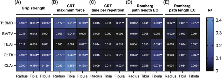
While osteoporosis was diagnosed in 115/230 (50.0%) women, sarcopenia was detected in 38/230 (16.5%). Positive associations of both grip strength and CRT maximum force with cortical geometric and microarchitectural parameters were detected at all measured sites, with the strongest effect applying to CRT maximum force and tibial parameters (e.g. tibial cortical area R = 0.36, P < 0.0001, and tibial cortical thickness R = 0.26, P < 0.0001). Balance parameters showed much weaker or no associations with HR-pQCT parameters. Major associations between muscle strength and trabecular parameters could not be confirmed. Age and body mass index were confirmed as negative and positive predictors for several microarchitectural parameters, respectively. An independent predictive value of grip strength on radial, tibial, and fibular (all P < 0.01) cortical area and of CRT maximum relative force on cortical thickness (all P < 0.05) was revealed. Women with osteosarcopenia showed significantly reduced cortical HR-pQCT parameters but no differences in DXA values compared with women with osteoporosis but no sarcopenia. Stratification by fracture and treatment status revealed that vertebral fractures and denosumab treatment altered the muscle-bone interaction.
A systemic interaction between muscle strength and bone microarchitecture was demonstrated, and this interaction appears to be primarily with the cortical bone compartment. The value of muscle assessments in fracture risk evaluation may be partly mediated by their effects on bone microarchitecture.
众所周知,骨骼完整性受肌肉影响。肌肉力量较弱(即肌肉减少症)被认为是脆性骨折的主要预测因素。虽然这一观察结果对于骨质疏松风险增加的老年女性尤为重要,但尚未有全面的研究来确定肌肉性能对多个身体部位特定部位骨微观结构的影响。
我们对 230 名(年龄 21 至 87 岁)高骨质疏松风险女性的不同肌肉性能和骨微观结构评估数据进行了回顾性分析。肌肉性能测试包括握力和坐起测试(CRT)以及肌动描记术。平衡通过 Romberg 姿势描记法确定。骨矿物质密度(BMD)通过髋部和腰椎的双能 X 射线吸收法(DXA)进行测量。通过第二代高分辨率外周定量 CT(HR-pQCT)在多个骨骼部位(桡骨远端、胫骨和腓骨)评估特定部位的容积 BMD、微观结构和几何形状。应用回归模型测试肌肉和骨骼参数之间的相互作用。根据骨质疏松症和骨质疏松-肌肉减少症的 BMD 和微观结构定义了亚组。
230 名女性中,115 名(50.0%)被诊断为骨质疏松症,38 名(16.5%)被诊断为肌肉减少症。在所有测量部位均发现握力和 CRT 最大力量与皮质几何和微观结构参数呈正相关,其中 CRT 最大力量和胫骨参数的相关性最强(例如,胫骨皮质面积 R = 0.36,P < 0.0001,和胫骨皮质厚度 R = 0.26,P < 0.0001)。平衡参数与 HR-pQCT 参数的相关性较弱或不存在。无法证实肌肉力量与松质骨参数之间存在主要关联。年龄和体重指数分别被证实为多个微观结构参数的负预测因子和正预测因子。握力对桡骨、胫骨和腓骨(均 P < 0.01)皮质面积和 CRT 最大相对力对皮质厚度(均 P < 0.05)的独立预测值。与骨质疏松症但无肌肉减少症的女性相比,患有骨质疏松-肌肉减少症的女性皮质 HR-pQCT 参数明显降低,但 DXA 值无差异。骨折和治疗状况的分层表明,椎体骨折和地舒单抗治疗改变了肌肉-骨骼相互作用。
证明了肌肉力量和骨微观结构之间存在系统相互作用,这种相互作用似乎主要与皮质骨有关。肌肉评估在骨折风险评估中的价值可能部分由其对骨微观结构的影响介导。


