• 文献检索
  • 文档翻译
  • 深度研究
  • 学术资讯
  • Suppr Zotero 插件Zotero 插件
  • 邀请有礼
  • 套餐&价格
  • 历史记录
应用&插件
Suppr Zotero 插件Zotero 插件浏览器插件Mac 客户端Windows 客户端微信小程序
定价
高级版会员购买积分包购买API积分包
服务
文献检索文档翻译深度研究API 文档MCP 服务
关于我们
关于 Suppr公司介绍联系我们用户协议隐私条款
关注我们

Suppr 超能文献

核心技术专利:CN118964589B侵权必究
粤ICP备2023148730 号-1Suppr @ 2026

文献检索

告别复杂PubMed语法,用中文像聊天一样搜索,搜遍4000万医学文献。AI智能推荐,让科研检索更轻松。

立即免费搜索

文件翻译

保留排版,准确专业,支持PDF/Word/PPT等文件格式,支持 12+语言互译。

免费翻译文档

深度研究

AI帮你快速写综述,25分钟生成高质量综述,智能提取关键信息,辅助科研写作。

立即免费体验

经支气管超声内镜针吸活检术(EBUS-TBNA)对原发性肺癌患者肺门、叶间和肺段淋巴结的临床应用价值。

Clinical utility of EBUS-TBNA of hilar, interlobar, and lobar lymph nodes in patients with primary lung cancer.

机构信息

Department of Internal Medicine, Seoul Medical Center, Seoul, Republic of Korea.

Division of Pulmonary and Critical Care Medicine, Department of Medicine, Samsung Medical Center, Sungkyunkwan University School of Medicine, Seoul, Republic of Korea.

出版信息

Thorac Cancer. 2022 Sep;13(17):2507-2514. doi: 10.1111/1759-7714.14587. Epub 2022 Jul 20.

DOI:10.1111/1759-7714.14587
PMID:35859356
原文链接:https://pmc.ncbi.nlm.nih.gov/articles/PMC9436691/
Abstract

BACKGROUND

Endobronchial ultrasound-guided transbronchial needle aspiration (EBUS-TBNA) is used to evaluate hilar/interlobar/lobar lymph nodes. This study aimed to assess the clinical utility of EBUS-TBNA for station 10/11/12 lymph nodes (LNs) in patients with primary lung cancer.

METHODS

This was a retrospective analysis of a prospectively collected database of patients with primary lung cancer who underwent EBUS-TBNA for station 10/11/12 LNs from January 2015 to December 2019. Patients with benign results from EBUS-TBNA who did not undergo surgical sampling/clinical follow-up or who received radiotherapy/chemotherapy were excluded.

RESULTS

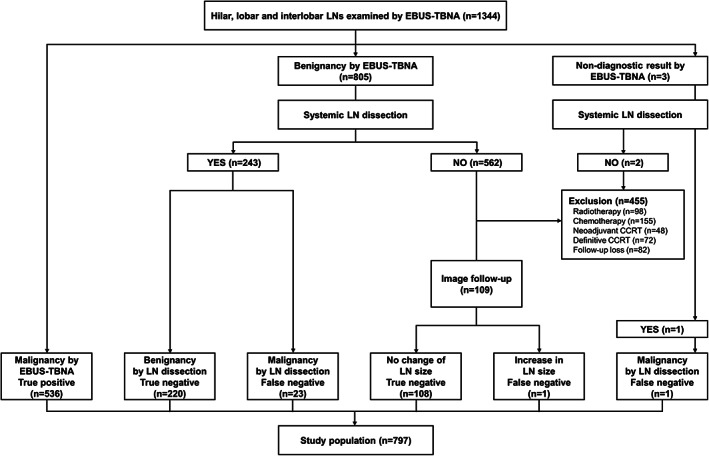
The analyses were conducted on 889 LNs from 797 patients. The overall diagnostic sensitivity, specificity, accuracy, negative predictive value (NPV), and positive predictive value of EBUS-TBNA were 95.7, 100, 97.3, 93.2, and 100%, respectively. Diagnostic sensitivity was significantly lower for LNs <10 mm than ≥10 mm in size (90.1% vs. 97.8%; p < 0.001). There was no significant difference in diagnostic performance according to the nodal station (10 vs. 11/12) and left- versus right-sided LNs. The diagnostic sensitivity (100 vs. 95.5%; p = 0.221) and specificity (100 vs. 100%) of N3 LNs was not significantly different from those of N1 LNs. In this study, eight (8/91, 8.8%) patients with cN1 NSCLC received neoadjuvant treatment based on the results of EBUS-TBNA.

CONCLUSION

EBUS-TBNA accurately evaluates station 10/11/12 LNs of both N1 and N3 disease. The diagnostic performances of EBUS-TBNA for station 10/11/12 LNs seem to be comparable to those of EBUS-TBNA for mediastinal LNs.

摘要

背景

经支气管超声引导针吸活检术(EBUS-TBNA)用于评估肺门/叶间/肺段淋巴结。本研究旨在评估 EBUS-TBNA 对原发性肺癌患者第 10/11/12 站淋巴结(LNs)的临床应用价值。

方法

这是一项对 2015 年 1 月至 2019 年 12 月期间接受 EBUS-TBNA 第 10/11/12 站 LN 活检的原发性肺癌患者前瞻性数据库的回顾性分析。EBUS-TBNA 结果为良性但未进行手术取样/临床随访或接受放疗/化疗的患者被排除在外。

结果

共对 797 例患者的 889 个 LN 进行了分析。EBUS-TBNA 的总体诊断敏感性、特异性、准确性、阴性预测值(NPV)和阳性预测值分别为 95.7%、100%、97.3%、93.2%和 100%。大小<10mm 的 LN 的诊断敏感性明显低于≥10mm 的 LN(90.1%比 97.8%;p<0.001)。根据淋巴结站(第 10 站与 11/12 站)和左/右侧 LN,诊断性能无显著差异。N3 LNs 的诊断敏感性(100%比 95.5%;p=0.221)和特异性(100%比 100%)与 N1 LNs 无显著差异。在本研究中,8 例(8/91,8.8%)cN1 NSCLC 患者根据 EBUS-TBNA 结果接受了新辅助治疗。

结论

EBUS-TBNA 可准确评估 N1 和 N3 疾病的第 10/11/12 站 LNs。EBUS-TBNA 对第 10/11/12 站 LNs 的诊断性能似乎与 EBUS-TBNA 对纵隔 LNs 的诊断性能相当。

https://cdn.ncbi.nlm.nih.gov/pmc/blobs/6db4/9436691/5e9eb900287a/TCA-13-2507-g001.jpg

相似文献

1
Clinical utility of EBUS-TBNA of hilar, interlobar, and lobar lymph nodes in patients with primary lung cancer.经支气管超声内镜针吸活检术(EBUS-TBNA)对原发性肺癌患者肺门、叶间和肺段淋巴结的临床应用价值。
Thorac Cancer. 2022 Sep;13(17):2507-2514. doi: 10.1111/1759-7714.14587. Epub 2022 Jul 20.
2
The critical role of EBUS-TBNA cytology in the staging of mediastinal lymph nodes in lung cancer patients: A correlation study with positron emission tomography findings.超声支气管镜引导下经支气管针吸活检术(EBUS-TBNA)细胞学检查在肺癌患者纵隔淋巴结分期中的关键作用:与正电子发射断层扫描结果的相关性研究
Cancer Cytopathol. 2017 Sep;125(9):717-725. doi: 10.1002/cncy.21886. Epub 2017 Jun 13.
3
Endobronchial ultrasound-guided transbronchial needle aspiration for differentiating N0 versus N1 lung cancer.经支气管超声引导针吸活检术用于区分 N0 与 N1 期肺癌。
Ann Thorac Surg. 2013 Nov;96(5):1756-60. doi: 10.1016/j.athoracsur.2013.05.090. Epub 2013 Aug 15.
4
The value of endobronchial ultrasound-guided transbronchial needle aspiration, 18-fluorodeoxyglucose positron emission tomography/computed tomography, and ultrasonography imaging techniques in the diagnosis of mediastinal and/or hilar malignant, anthracotic, and other benign lymph nodes.支气管内超声引导下经支气管针吸活检、18氟脱氧葡萄糖正电子发射断层扫描/计算机断层扫描及超声成像技术在纵隔和/或肺门恶性、炭末沉着性及其他良性淋巴结诊断中的价值
Medicine (Baltimore). 2021 Feb 19;100(7):e24728. doi: 10.1097/MD.0000000000024728.
5
The Utility of Endobronchial Ultrasound-guided Transbronchial Needle Aspiration in Patients with Small-cell Lung Cancer.支气管内超声引导下经支气管针吸活检术在小细胞肺癌患者中的应用价值
Intern Med. 2016;55(9):1061-6. doi: 10.2169/internalmedicine.55.6082. Epub 2016 May 1.
6
Nodal stations and diagnostic performances of endobronchial ultrasound-guided transbronchial needle aspiration in patients with non-small cell lung cancer.经支气管超声引导针吸活检术在非小细胞肺癌患者中的淋巴结站和诊断性能。
J Korean Med Sci. 2012 Jan;27(1):46-51. doi: 10.3346/jkms.2012.27.1.46. Epub 2011 Dec 19.
7
Is it necessary to sample the contralateral nodal stations by EBUS-TBNA in patients with lung cancer and clinical N0 / N1 on PET-CT?在 PET-CT 临床 N0/N1 肺癌患者中,是否有必要通过 EBUS-TBNA 对侧淋巴结站取样?
Lung Cancer. 2020 Apr;142:9-12. doi: 10.1016/j.lungcan.2020.01.014. Epub 2020 Jan 13.
8
Quantitative analysis of endobronchial ultrasound elastography in computed tomography-negative mediastinal and hilar lymph nodes.经 CT 检查为阴性的纵隔和肺门淋巴结的支气管内超声弹性成像的定量分析。
Thorac Cancer. 2020 Sep;11(9):2590-2599. doi: 10.1111/1759-7714.13579. Epub 2020 Jul 21.
9
Impact of F-FDG PET/CT, CT and EBUS/TBNA on preoperative mediastinal nodal staging of NSCLC.18F-氟脱氧葡萄糖正电子发射断层扫描/计算机断层扫描、计算机断层扫描和支气管内超声引导针吸活检术对非小细胞肺癌术前纵隔淋巴结分期的影响。
BMC Med Imaging. 2021 Mar 17;21(1):49. doi: 10.1186/s12880-021-00580-w.
10
Combined endobronchial and esophageal endosonography for the diagnosis and staging of lung cancer: European Society of Gastrointestinal Endoscopy (ESGE) Guideline, in cooperation with the European Respiratory Society (ERS) and the European Society of Thoracic Surgeons (ESTS).支气管内与食管内超声联合用于肺癌诊断及分期:欧洲胃肠内镜学会(ESGE)指南,与欧洲呼吸学会(ERS)及欧洲胸外科医师学会(ESTS)合作制定
Endoscopy. 2015 Jun;47(6):545-59. doi: 10.1055/s-0034-1392040. Epub 2015 Jun 1.

引用本文的文献

1
Portrayal of video-assisted mediastinoscopic lymphadenectomy's range subsequent to its simultaneous use with uniportal VAT-lobectomy for left-sided NSCLC: a case-based perspective.在左侧非小细胞肺癌的单孔胸腔镜解剖性肺段切除术(VAT-lobectomy)中同时使用电视辅助纵隔镜淋巴结清扫术的范围:基于病例的观点。
J Cardiothorac Surg. 2023 Apr 17;18(1):152. doi: 10.1186/s13019-023-02277-3.
2
Diagnostic Performance of Endosonography to Detect Mediastinal Lymph Node Metastasis in Patients with Radiological N1 Non-Small Cell Lung Cancer.超声内镜诊断影像学 N1 期非小细胞肺癌纵隔淋巴结转移的效能。
Cancer Res Treat. 2023 Jul;55(3):832-840. doi: 10.4143/crt.2022.1428. Epub 2023 Mar 2.

本文引用的文献

1
Clinical Usefulness of Fungal Culture of EBUS-TBNA Needle Rinse Fluid and Core Tissue.经支气管超声引导针吸活检术(EBUS-TBNA)针冲洗液和核心组织真菌培养的临床应用
Yonsei Med J. 2020 Aug;61(8):670-678. doi: 10.3349/ymj.2020.61.8.670.
2
Cancer statistics, 2020.癌症统计数据,2020 年。
CA Cancer J Clin. 2020 Jan;70(1):7-30. doi: 10.3322/caac.21590. Epub 2020 Jan 8.
3
The utility of endosonography for mediastinal staging of non-small cell lung cancer in patients with radiological N0 disease.超声内镜对于影像学 N0 期非小细胞肺癌患者纵隔分期的作用。
Lung Cancer. 2020 Jan;139:151-156. doi: 10.1016/j.lungcan.2019.11.021. Epub 2019 Nov 26.
4
Relationship between endobronchial ultrasound-guided (EBUS)-transbronchial needle aspiration utility and computed tomography staging, node size at EBUS, and positron emission tomography scan node standard uptake values: A retrospective analysis.经支气管超声引导(EBUS)-经支气管针吸术的应用与计算机断层扫描分期、EBUS 时的淋巴结大小和正电子发射断层扫描(PET)扫描淋巴结标准摄取值之间的关系:一项回顾性分析。
Thorac Cancer. 2017 Jul;8(4):285-290. doi: 10.1111/1759-7714.12438. Epub 2017 Apr 24.
5
The International Association for the Study of Lung Cancer Lung Cancer Staging Project: Proposals for the Revision of the N Descriptors in the Forthcoming 8th Edition of the TNM Classification for Lung Cancer.国际肺癌研究协会肺癌分期项目:对即将发布的第 8 版肺癌 TNM 分类中 N 描述符修订的建议。
J Thorac Oncol. 2015 Dec;10(12):1675-84. doi: 10.1097/JTO.0000000000000678.
6
Combined endobronchial and esophageal endosonography for the diagnosis and staging of lung cancer: European Society of Gastrointestinal Endoscopy (ESGE) Guideline, in cooperation with the European Respiratory Society (ERS) and the European Society of Thoracic Surgeons (ESTS).支气管内与食管内超声联合用于肺癌诊断及分期:欧洲胃肠内镜学会(ESGE)指南,与欧洲呼吸学会(ERS)及欧洲胸外科医师学会(ESTS)合作制定
Endoscopy. 2015 Jun;47(6):545-59. doi: 10.1055/s-0034-1392040. Epub 2015 Jun 1.
7
Diagnosing and staging lung cancer involving the mediastinum.诊断和分期累及纵隔的肺癌。
Chest. 2015 May;147(5):1401-1412. doi: 10.1378/chest.14-1355.
8
Endobronchial ultrasound versus mediastinoscopy for mediastinal nodal staging of non-small-cell lung cancer.经支气管超声内镜与纵隔镜检查用于非小细胞肺癌纵隔淋巴结分期。
J Thorac Oncol. 2015 Feb;10(2):331-7. doi: 10.1097/JTO.0000000000000388.
9
Revised ESTS guidelines for preoperative mediastinal lymph node staging for non-small-cell lung cancer.非小细胞肺癌术前纵隔淋巴结分期的修订版欧洲胸外科医师协会指南。
Eur J Cardiothorac Surg. 2014 May;45(5):787-98. doi: 10.1093/ejcts/ezu028. Epub 2014 Feb 26.
10
Survival of patients with unsuspected pN2 non-small cell lung cancer after an accurate preoperative mediastinal staging.术前纵隔准确分期后未被怀疑的 pN2 非小细胞肺癌患者的生存情况。
Ann Thorac Surg. 2014 Mar;97(3):957-64. doi: 10.1016/j.athoracsur.2013.09.101. Epub 2013 Nov 26.