School of Biomedicine, Robinson Research Institute, The University of Adelaide, Adelaide, South Australia, Australia.
Australian Research Council Centre of Excellence for Nanoscale Biophotonics, The University of Adelaide, Adelaide, South Australia, Australia.
Biol Reprod. 2022 Oct 11;107(4):1014-1025. doi: 10.1093/biolre/ioac145.
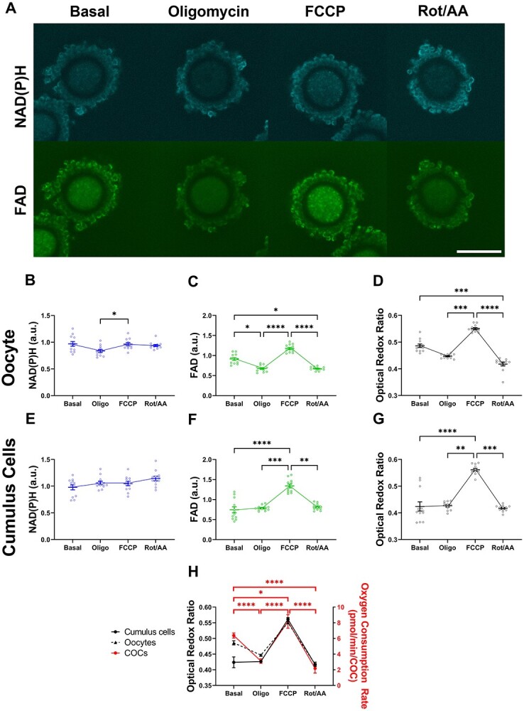
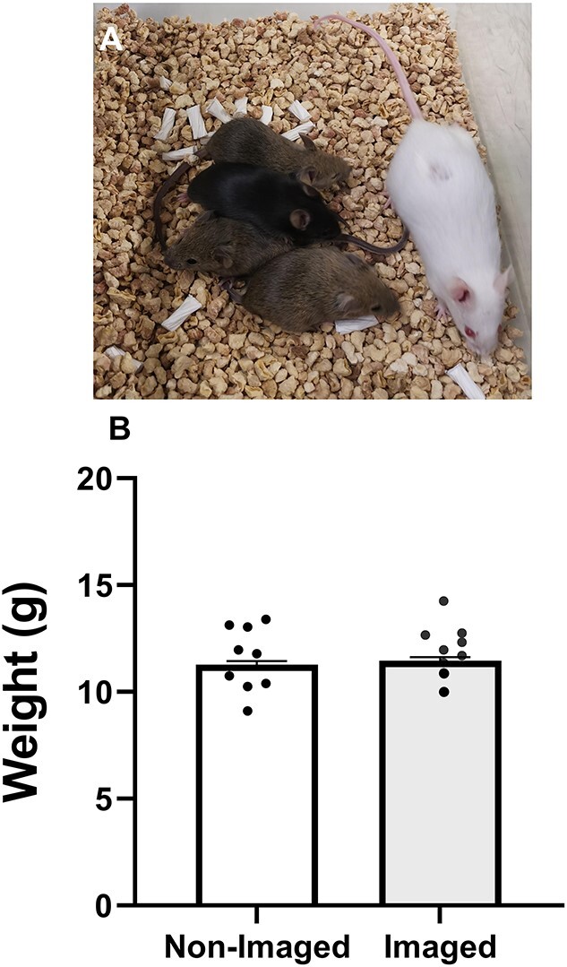
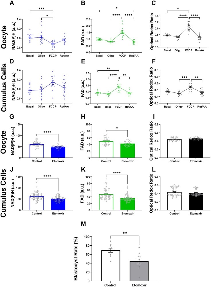
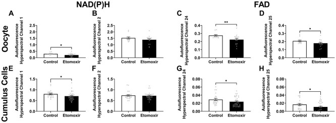
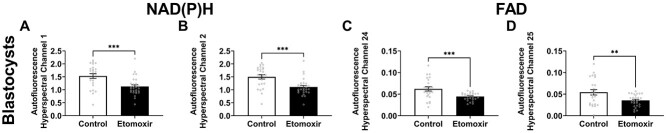
Oocyte developmental potential is intimately linked to metabolism. Existing approaches to measure metabolism in the cumulus oocyte complex (COC) do not provide information on the separate cumulus and oocyte compartments. Development of an assay that achieves this may lead to an accurate diagnostic for oocyte quality. Optical imaging of the autofluorescent cofactors reduced nicotinamide adenine dinucleotide (phosphate) [NAD(P)H] and flavin adenine dinucleotide (FAD) provides a spatially resolved indicator of metabolism via the optical redox ratio (FAD/[NAD(P)H + FAD]). This may provide an assessment of oocyte quality. Here, we determined whether the optical redox ratio is a robust methodology for measuring metabolism in the cumulus and oocyte compartments compared with oxygen consumption in the whole COC. We also determined whether optical imaging could detect metabolic differences associated with poor oocyte quality (etomoxir-treated). We used confocal microscopy to measure NAD(P)H and FAD, and extracellular flux to measure oxygen consumption. The optical redox ratio accurately reflected metabolism in the oocyte compartment when compared with oxygen consumption (whole COC). Etomoxir-treated COCs showed significantly lower levels of NAD(P)H and FAD compared to control. We further validated this approach using hyperspectral imaging, which is clinically compatible due to its low energy dose. This confirmed lower NAD(P)H and FAD in etomoxir-treated COCs. When comparing hyperspectral imaged vs non-imaged COCs, subsequent preimplantation development and post-transfer viability were comparable. Collectively, these results demonstrate that label-free optical imaging of metabolic cofactors is a safe and sensitive assay for measuring metabolism and has potential to assess oocyte developmental competence.
卵母细胞的发育潜能与代谢密切相关。现有的测量卵丘-卵母细胞复合物(COC)代谢的方法不能提供关于分开的卵丘和卵母细胞区室的信息。开发一种能够实现这一目标的测定方法可能会为卵母细胞质量提供一个准确的诊断。通过光学氧化还原比(FAD/[NAD(P)H + FAD])对还原型烟酰胺腺嘌呤二核苷酸(磷酸)[NAD(P)H]和黄素腺嘌呤二核苷酸(FAD)这两种自发荧光辅助因子进行光学成像,为通过光学方法测量代谢提供了一种具有空间分辨率的指示剂。这可能为卵母细胞质量评估提供一种手段。在这里,我们确定了与整个 COC 的耗氧量相比,光学氧化还原比是否是一种测量卵丘和卵母细胞区室代谢的可靠方法。我们还确定了光学成像是否可以检测与卵母细胞质量差(使用 etomoxir 处理)相关的代谢差异。我们使用共聚焦显微镜测量 NAD(P)H 和 FAD,以及使用细胞外通量测量耗氧量。与耗氧量(整个 COC)相比,光学氧化还原比准确反映了卵母细胞区室的代谢情况。与对照相比,用 etomoxir 处理的 COC 中 NAD(P)H 和 FAD 的水平明显降低。我们使用高光谱成像进一步验证了这种方法,由于其低能量剂量,该方法在临床上是兼容的。这证实了用 etomoxir 处理的 COC 中 NAD(P)H 和 FAD 的含量较低。当比较高光谱成像和非成像 COC 时,随后的胚胎植入前发育和移植后活力相当。总之,这些结果表明,代谢辅助因子的无标记光学成像方法是一种安全且敏感的测量代谢的方法,并且有可能评估卵母细胞的发育能力。