Robinson Research Institute, School of Biomedicine, The University of Adelaide, Adelaide, Australia.
Institute for Photonics and Advanced Sensing, The University of Adelaide, Adelaide, Australia.
Sci Rep. 2024 Sep 5;14(1):20760. doi: 10.1038/s41598-024-71443-x.
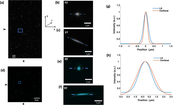
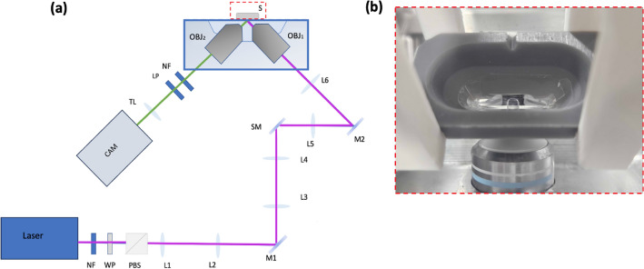
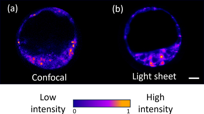
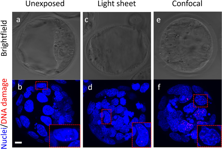
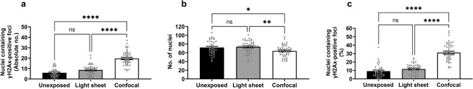
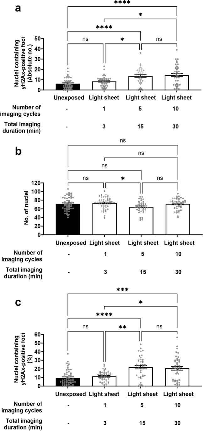
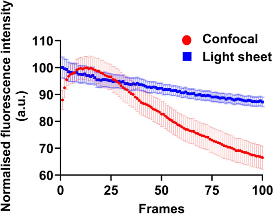
Embryo quality assessment by optical imaging is increasing in popularity. Among available optical techniques, light sheet microscopy has emerged as a superior alternative to confocal microscopy due to its geometry, enabling faster image acquisition with reduced photodamage to the sample. However, previous assessments of photodamage induced by imaging may have failed to measure more subtle impacts. In this study, we employed DNA damage as a sensitive indicator of photodamage. We use light sheet microscopy with excitation at a wavelength of 405 nm for imaging embryo autofluorescence and compare its performance to laser scanning confocal microscopy. At an equivalent signal-to-noise ratio for images acquired with both modalities, light sheet microscopy reduced image acquisition time by ten-fold, and did not induce DNA damage when compared to non-imaged embryos. In contrast, imaging with confocal microscopy led to significantly higher levels of DNA damage within embryos and had a higher photobleaching rate. Light sheet imaging is also capable of inducing DNA damage within the embryo but requires multiple cycles of volumetric imaging. Collectively, this study confirms that light sheet microscopy is faster and safer than confocal microscopy for imaging live embryos, indicating its potential as a label-free diagnostic for embryo quality.
胚胎质量的光学成像评估越来越受到关注。在现有的光学技术中,由于其独特的几何结构,光片显微镜作为共聚焦显微镜的一种替代方法已经脱颖而出,能够以更低的光损伤实现更快的图像采集。然而,以前对成像引起的光损伤的评估可能未能测量到更细微的影响。在这项研究中,我们将 DNA 损伤作为光损伤的敏感指标。我们使用波长为 405nm 的光片显微镜对胚胎自发荧光进行成像,并将其性能与激光扫描共聚焦显微镜进行比较。在两种模式下获取的图像具有相同的信噪比时,光片显微镜将图像采集时间缩短了十倍,并且与非成像胚胎相比不会引起 DNA 损伤。相比之下,共聚焦显微镜成像会导致胚胎内的 DNA 损伤显著增加,并且具有更高的光漂白率。光片成像也有能力在胚胎内诱导 DNA 损伤,但需要多次体积成像循环。总的来说,这项研究证实了光片显微镜在对活胚胎进行成像时比共聚焦显微镜更快、更安全,表明其有潜力成为胚胎质量的无标记诊断方法。