Departments of Cardiovascular Medicine.
Radiology Laboratory, Fujiikai Kashibaseiki Hospital, Kashiba.
Coron Artery Dis. 2022 Nov 1;33(7):531-539. doi: 10.1097/MCA.0000000000001171. Epub 2022 Jul 22.
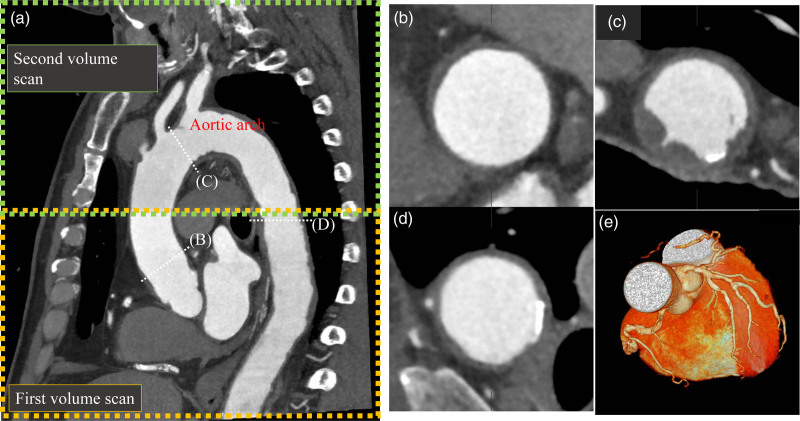
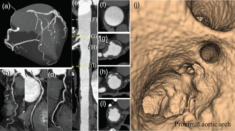
Wide-volume scanning with 320-row multidetector computed tomography coronary angiography (CTCA-WVS) enables the assessment of the aortic arch plaque (AAP) morphology and coronary arteries without requiring additional contrast volume. This study aimed to investigate the prevalence of AAPs and their association with coronary artery disease (CAD) and major adverse cardiovascular events (MACEs) in patients who underwent CTCA-WVS.
This study included 204 patients without known CAD (mean age, 65 years; 53% men) who underwent CTCA-WVS. We evaluated the presence of aortic plaques in the ascending aorta, aortic arch, and thoracic descending aorta using CTCA-WVS. Large aortic plaques were defined as plaques of at least 4 mm in thickness. A complex aortic plaque was defined as a plaque with ulceration or protrusion. MACEs were defined as composite events of cardiovascular (CV) death, nonfatal myocardial infarction, and ischemic stroke.
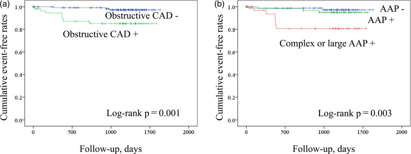
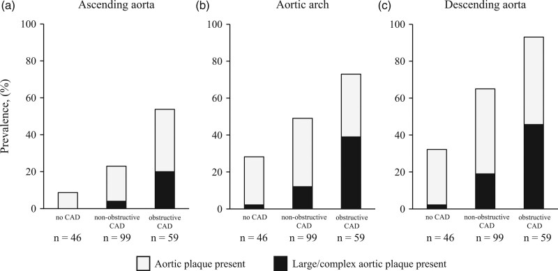
AAPs and large/complex AAPs were identified in 51% ( n = 105) and 18% ( n = 36) of the study patients, respectively. The prevalence of AAPs with large/complex morphology increased with CAD severity (2.1% in no CAD, 12% in nonobstructive CAD, and 39% in obstructive CAD). The univariate Cox hazard model demonstrated that the predictors associated with MACEs were diabetes, obstructive CAD, and large/complex AAPs. Independent factors associated with large/complex AAPs were male sex [odds ratio (OR), 2.90; P = 0.025], stroke history (OR, 3.48; P = 0.026), obstructive CAD (OR, 3.35; P = 0.011), and thoracic aortic calcification (OR, 1.77; P = 0.005).
CTCA-WVS provides a comprehensive assessment of coronary atherosclerosis and thoracic aortic plaques in patients with CAD, which may improve the stratification of patients at risk for CV events.
320 排多层螺旋 CT 冠状动脉血管成像(CTCA-WVS)大容积扫描可在不增加对比剂用量的情况下评估主动脉弓斑块(AAP)形态和冠状动脉,本研究旨在探讨 CTCA-WVS 检查中 AAP 的发生率及其与冠状动脉疾病(CAD)和主要不良心血管事件(MACE)的相关性。
本研究纳入了 204 例无已知 CAD(平均年龄 65 岁,53%为男性)的患者,所有患者均接受 CTCA-WVS 检查。我们使用 CTCA-WVS 评估升主动脉、主动脉弓和胸降主动脉中主动脉斑块的存在。大的主动脉斑块定义为厚度至少 4mm 的斑块。复杂的主动脉斑块定义为溃疡或突起的斑块。MACE 定义为心血管(CV)死亡、非致死性心肌梗死和缺血性卒中的复合事件。
51%(n=105)和 18%(n=36)的研究患者存在 AAP 和大/复杂 AAP。AAP 伴有大/复杂形态的发生率随着 CAD 严重程度的增加而增加(无 CAD 患者为 2.1%,非阻塞性 CAD 患者为 12%,阻塞性 CAD 患者为 39%)。单变量 Cox 风险模型显示,与 MACE 相关的预测因素为糖尿病、阻塞性 CAD 和大/复杂 AAP。与大/复杂 AAP 相关的独立因素为男性(优势比[OR],2.90;P=0.025)、卒中史(OR,3.48;P=0.026)、阻塞性 CAD(OR,3.35;P=0.011)和胸主动脉钙化(OR,1.77;P=0.005)。
CTCA-WVS 为 CAD 患者提供了冠状动脉粥样硬化和胸主动脉斑块的全面评估,这可能有助于对 CV 事件风险患者进行分层。