• 文献检索
  • 文档翻译
  • 深度研究
  • 学术资讯
  • Suppr Zotero 插件Zotero 插件
  • 邀请有礼
  • 套餐&价格
  • 历史记录
应用&插件
Suppr Zotero 插件Zotero 插件浏览器插件Mac 客户端Windows 客户端微信小程序
定价
高级版会员购买积分包购买API积分包
服务
文献检索文档翻译深度研究API 文档MCP 服务
关于我们
关于 Suppr公司介绍联系我们用户协议隐私条款
关注我们

Suppr 超能文献

核心技术专利:CN118964589B侵权必究
粤ICP备2023148730 号-1Suppr @ 2026

文献检索

告别复杂PubMed语法,用中文像聊天一样搜索,搜遍4000万医学文献。AI智能推荐,让科研检索更轻松。

立即免费搜索

文件翻译

保留排版,准确专业,支持PDF/Word/PPT等文件格式,支持 12+语言互译。

免费翻译文档

深度研究

AI帮你快速写综述,25分钟生成高质量综述,智能提取关键信息,辅助科研写作。

立即免费体验

开发改良的 Lats 激酶抑制剂以促进哺乳动物器官的再生。

Development of an improved inhibitor of Lats kinases to promote regeneration of mammalian organs.

机构信息

Howard Hughes Medical Institute and Laboratory of Sensory Neuroscience, The Rockefeller University, New York, NY 10065.

Tri-Institutional Therapeutics Discovery Institute, New York, NY 10021.

出版信息

Proc Natl Acad Sci U S A. 2022 Jul 12;119(28):e2206113119. doi: 10.1073/pnas.2206113119. Epub 2022 Jul 8.

DOI:10.1073/pnas.2206113119
PMID:35867764
原文链接:https://pmc.ncbi.nlm.nih.gov/articles/PMC9282237/
Abstract

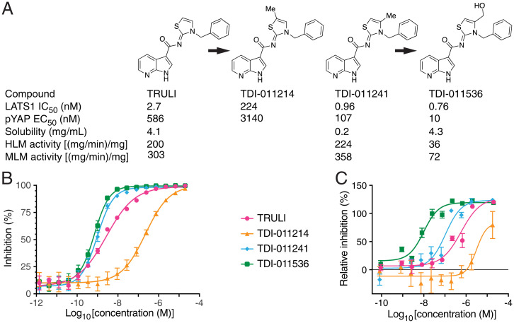
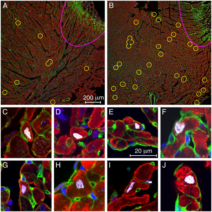
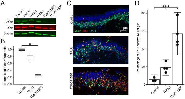
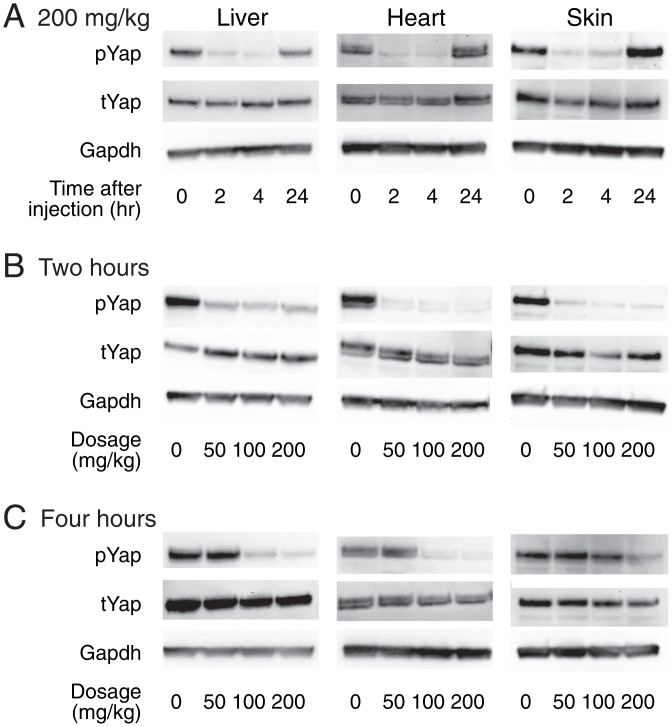
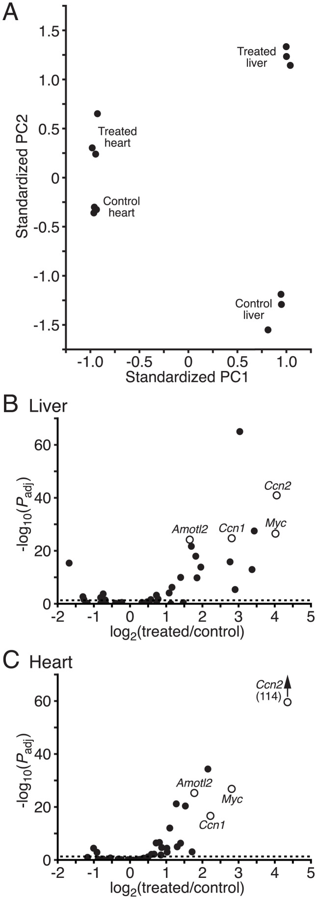
The Hippo signaling pathway acts as a brake on regeneration in many tissues. This cascade of kinases culminates in the phosphorylation of the transcriptional cofactors Yap and Taz, whose concentration in the nucleus consequently remains low. Various types of cellular signals can reduce phosphorylation, however, resulting in the accumulation of Yap and Taz in the nucleus and subsequently in mitosis. We earlier identified a small molecule, TRULI, that blocks the final kinases in the pathway, Lats1 and Lats2, and thus elicits proliferation of several cell types that are ordinarily postmitotic and aids regeneration in mammals. In the present study, we present the results of chemical modification of the original compound and demonstrate that a derivative, TDI-011536, is an effective blocker of Lats kinases in vitro at nanomolar concentrations. The compound fosters extensive proliferation in retinal organoids derived from human induced pluripotent stem cells. Intraperitoneal administration of the substance to mice suppresses Yap phosphorylation for several hours and induces transcriptional activation of Yap target genes in the heart, liver, and skin. Moreover, the compound initiates the proliferation of cardiomyocytes in adult mice following cardiac cryolesions. After further chemical refinement, related compounds might prove useful in protective and regenerative therapies.

摘要

Hippo 信号通路在许多组织的再生中起制动作用。这一系列激酶最终导致转录共激活因子 Yap 和 Taz 的磷酸化,其核内浓度因此保持较低水平。然而,各种类型的细胞信号可以减少磷酸化,导致 yap 和 Taz 在核内积累,随后进入有丝分裂。我们之前鉴定出一种小分子 TRULI,它可以阻断通路中的最后两种激酶 Lats1 和 Lats2,从而引发几种通常处于有丝分裂后状态的细胞类型的增殖,并有助于哺乳动物的再生。在本研究中,我们介绍了对原始化合物进行化学修饰的结果,并证明一种衍生物 TDI-011536 在纳摩尔浓度下是 Lats 激酶的有效抑制剂。该化合物可促进源自人诱导多能干细胞的视网膜类器官的广泛增殖。该物质通过腹腔给药可在数小时内抑制 Yap 的磷酸化,并在心脏、肝脏和皮肤中诱导 yap 靶基因的转录激活。此外,该化合物在心脏冷冻损伤后可引发成年小鼠心肌细胞的增殖。经过进一步的化学优化,相关化合物可能在保护和再生治疗中具有应用价值。

https://cdn.ncbi.nlm.nih.gov/pmc/blobs/3166/9282237/98a7eb2b0d63/pnas.2206113119fig05.jpg
https://cdn.ncbi.nlm.nih.gov/pmc/blobs/3166/9282237/65c9cfc0ea8d/pnas.2206113119fig01.jpg
https://cdn.ncbi.nlm.nih.gov/pmc/blobs/3166/9282237/1f38cb075f75/pnas.2206113119fig02.jpg
https://cdn.ncbi.nlm.nih.gov/pmc/blobs/3166/9282237/23ff51e78fdf/pnas.2206113119fig03.jpg
https://cdn.ncbi.nlm.nih.gov/pmc/blobs/3166/9282237/798f8d35c24a/pnas.2206113119fig04.jpg
https://cdn.ncbi.nlm.nih.gov/pmc/blobs/3166/9282237/98a7eb2b0d63/pnas.2206113119fig05.jpg

相似文献

1
Development of an improved inhibitor of Lats kinases to promote regeneration of mammalian organs.开发改良的 Lats 激酶抑制剂以促进哺乳动物器官的再生。
Proc Natl Acad Sci U S A. 2022 Jul 12;119(28):e2206113119. doi: 10.1073/pnas.2206113119. Epub 2022 Jul 8.
2
NIBR-LTSi is a selective LATS kinase inhibitor activating YAP signaling and expanding tissue stem cells in vitro and in vivo.NIBR-LTSi 是一种选择性的 LATS 激酶抑制剂,可激活 YAP 信号通路,并在体外和体内扩增组织干细胞。
Cell Stem Cell. 2024 Apr 4;31(4):554-569.e17. doi: 10.1016/j.stem.2024.03.003.
3
Small-molecule inhibition of Lats kinases may promote Yap-dependent proliferation in postmitotic mammalian tissues.小分子抑制 Lats 激酶可能会促进有丝分裂后哺乳动物组织中 Yap 依赖性的增殖。
Nat Commun. 2021 May 25;12(1):3100. doi: 10.1038/s41467-021-23395-3.
4
YAP Mediates Hair Cell Regeneration in Balance Organs of Chickens, But LATS Kinases Suppress Its Activity in Mice.YAP 在鸡的平衡器官中介导毛细胞再生,但 LATS 激酶在小鼠中抑制其活性。
J Neurosci. 2020 May 13;40(20):3915-3932. doi: 10.1523/JNEUROSCI.0306-20.2020. Epub 2020 Apr 27.
5
Tudor-SN promotes cardiomyocyte proliferation and neonatal heart regeneration through regulating the phosphorylation of YAP.Tudor-SN 通过调节 YAP 的磷酸化促进心肌细胞增殖和新生心脏再生。
Cell Commun Signal. 2024 Jun 28;22(1):345. doi: 10.1186/s12964-024-01715-6.
6
WWC1 and NF2 Prevent the Development of Intrahepatic Cholangiocarcinoma by Regulating YAP/TAZ Activity through LATS in Mice.WWC1 和 NF2 通过调节 LATS 在小鼠中抑制 YAP/TAZ 活性从而预防肝内胆管细胞癌的发生。
Mol Cells. 2020 May 31;43(5):491-499. doi: 10.14348/molcells.2020.0093.
7
Large tumor suppressor homologs 1 and 2 regulate mouse liver progenitor cell proliferation and maturation through antagonism of the coactivators YAP and TAZ.大肿瘤抑制同源物1和2通过拮抗共激活因子YAP和TAZ来调节小鼠肝祖细胞的增殖和成熟。
Hepatology. 2016 Nov;64(5):1757-1772. doi: 10.1002/hep.28768. Epub 2016 Sep 30.
8
Deubiquitinating Enzyme USP9X Suppresses Tumor Growth via LATS Kinase and Core Components of the Hippo Pathway.去泛素化酶USP9X通过LATS激酶和Hippo信号通路的核心组件抑制肿瘤生长。
Cancer Res. 2017 Sep 15;77(18):4921-4933. doi: 10.1158/0008-5472.CAN-16-3413. Epub 2017 Jul 18.
9
Sphingosylphosphorylcholine regulates the Hippo signaling pathway in a dual manner.鞘氨醇磷酸胆碱以双重方式调节Hippo信号通路。
Cell Signal. 2016 Dec;28(12):1894-1903. doi: 10.1016/j.cellsig.2016.09.004. Epub 2016 Sep 12.
10
Upstream regulation of the Hippo-Yap pathway in cardiomyocyte regeneration.心肌细胞再生中的 Hippo-Yap 通路的上游调控。
Semin Cell Dev Biol. 2020 Apr;100:11-19. doi: 10.1016/j.semcdb.2019.09.004. Epub 2019 Oct 9.

引用本文的文献

1
Discovery and Optimization of LATS1 and LATS2 Kinase Inhibitors for Use in Regenerative Medicine.用于再生医学的LATS1和LATS2激酶抑制剂的发现与优化
J Med Chem. 2025 Aug 28;68(16):17499-17515. doi: 10.1021/acs.jmedchem.5c01027. Epub 2025 Aug 6.
2
Identifying the Role of YAP in the Development of Rumen Epithelium Using 3D Organoid.利用三维类器官鉴定YAP在瘤胃上皮发育中的作用
Stem Cells Int. 2025 Jul 11;2025:5105796. doi: 10.1155/sci/5105796. eCollection 2025.
3
Targeting the Hippo pathway in cancer.靶向癌症中的Hippo信号通路。

本文引用的文献

1
Small molecule LATS kinase inhibitors block the Hippo signaling pathway and promote cell growth under 3D culture conditions.小分子 LATS 激酶抑制剂在 3D 培养条件下阻断 Hippo 信号通路并促进细胞生长。
J Biol Chem. 2022 Apr;298(4):101779. doi: 10.1016/j.jbc.2022.101779. Epub 2022 Feb 26.
2
Small-molecule inhibition of Lats kinases may promote Yap-dependent proliferation in postmitotic mammalian tissues.小分子抑制 Lats 激酶可能会促进有丝分裂后哺乳动物组织中 Yap 依赖性的增殖。
Nat Commun. 2021 May 25;12(1):3100. doi: 10.1038/s41467-021-23395-3.
3
The Effects of YAP and Its Related Mechanisms in Central Nervous System Diseases.
Nat Rev Drug Discov. 2025 Jun 30. doi: 10.1038/s41573-025-01234-0.
4
Small-Molecule Inhibition of the Hippo Pathway Induces Regeneration of Retinal Pigment Epithelium.小分子抑制Hippo信号通路可诱导视网膜色素上皮细胞再生。
Invest Ophthalmol Vis Sci. 2025 Jun 2;66(6):69. doi: 10.1167/iovs.66.6.69.
5
A Universal 6iL/E4 Culture System for Deriving and Maintaining Embryonic Stem Cells Across Mammalian Species.一种用于衍生和维持跨哺乳动物物种胚胎干细胞的通用6iL/E4培养系统。
bioRxiv. 2025 May 21:2025.05.20.654948. doi: 10.1101/2025.05.20.654948.
6
Disrupting bile acid metabolism by suppressing Fxr causes hepatocellular carcinoma induced by YAP activation.通过抑制法尼醇X受体(Fxr)破坏胆汁酸代谢会导致由Yes相关蛋白(YAP)激活诱导的肝细胞癌。
Nat Commun. 2025 Apr 15;16(1):3583. doi: 10.1038/s41467-025-58809-z.
7
The Hippo pathway and p27 cooperate to suppress mitotic regeneration in the organ of Corti and the retina.河马通路和p27协同作用,抑制柯蒂氏器和视网膜中的有丝分裂再生。
Proc Natl Acad Sci U S A. 2025 Apr 8;122(14):e2411313122. doi: 10.1073/pnas.2411313122. Epub 2025 Apr 3.
8
LATS1-modulated ZBTB20 perturbing cartilage matrix homeostasis contributes to early-stage osteoarthritis.LATS1调节的ZBTB20破坏软骨基质稳态促成早期骨关节炎。
Bone Res. 2025 Mar 12;13(1):33. doi: 10.1038/s41413-025-00414-3.
9
Model construction and clinical therapeutic potential of engineered cardiac organoids for cardiovascular diseases.用于心血管疾病的工程化心脏类器官的模型构建及临床治疗潜力
Biomater Transl. 2024 Nov 15;5(4):337-354. doi: 10.12336/biomatertransl.2024.04.002. eCollection 2024.
10
Transient proliferation by reversible YAP and mitogen-control of the cyclin D1/p27 ratio.通过可逆的YAP实现瞬时增殖以及细胞周期蛋白D1/p27比率的有丝分裂原调控。
bioRxiv. 2024 Nov 4:2024.10.11.617852. doi: 10.1101/2024.10.11.617852.
YAP在中枢神经系统疾病中的作用及其相关机制
Front Neurosci. 2020 Jun 26;14:595. doi: 10.3389/fnins.2020.00595. eCollection 2020.
4
The regulation of Yorkie, YAP and TAZ: new insights into the Hippo pathway.Yorkie、YAP 和 TAZ 的调控:Hippo 通路的新见解。
Development. 2020 Apr 27;147(8):dev179069. doi: 10.1242/dev.179069.
5
Direct Comparison of Mononucleated and Binucleated Cardiomyocytes Reveals Molecular Mechanisms Underlying Distinct Proliferative Competencies.单核与双核心肌细胞的直接比较揭示了不同增殖能力背后的分子机制。
Cell Rep. 2020 Mar 3;30(9):3105-3116.e4. doi: 10.1016/j.celrep.2020.02.034.
6
Identification of Quinolinols as Activators of TEAD-Dependent Transcription.鉴定喹啉醇类化合物为 TEAD 依赖性转录的激活剂。
ACS Chem Biol. 2019 Dec 20;14(12):2909-2921. doi: 10.1021/acschembio.9b00786. Epub 2019 Dec 2.
7
Hippo-YAP/TAZ signalling in organ regeneration and regenerative medicine.Hippo-YAP/TAZ 信号通路在器官再生和再生医学中的作用。
Nat Rev Mol Cell Biol. 2019 Apr;20(4):211-226. doi: 10.1038/s41580-018-0086-y.
8
Mediated nuclear import and export of TAZ and the underlying molecular requirements.TAZ 的中介核输入和输出及潜在的分子需求。
Nat Commun. 2018 Nov 23;9(1):4966. doi: 10.1038/s41467-018-07450-0.
9
YAP/TAZ upstream signals and downstream responses.YAP/TAZ 的上游信号和下游反应。
Nat Cell Biol. 2018 Aug;20(8):888-899. doi: 10.1038/s41556-018-0142-z. Epub 2018 Jul 26.
10
Quantitative Analysis Reveals that Actin and Src-Family Kinases Regulate Nuclear YAP1 and Its Export.定量分析揭示肌动蛋白和Src 家族激酶调控核 YAP1 及其输出。
Cell Syst. 2018 Jun 27;6(6):692-708.e13. doi: 10.1016/j.cels.2018.05.006. Epub 2018 Jun 18.