Ariello Leonardo E, Mello Luiz Guilherme Marchesi, Pimentel Sérgio Luis Gianotti, Monteiro Mário L R
Division of Ophthalmology, University of São Paulo Medical School (USP), Av. Dr Enéas de Carvalho Aguiar, 255, Cerqueira César, São Paulo, SP, 05403-001, Brazil.
Departamento de Medicina Especializada, Federal University of Espírito Santo, Espírito Santo, Brazil.
Int J Retina Vitreous. 2022 Jul 22;8(1):48. doi: 10.1186/s40942-022-00403-2.
Papilledema is the main ocular finding in patients with idiopathic intracranial hypertension (IIH) although several chorioretinal abnormalities may also occur and contribute to visual loss. The purpose of this paper is to describe two cases of chorioretinal abnormalities associated with idiopathic intracranial hypertension: one with choroidal folds and another with polypoidal choroidal vasculopathy, an extremely unusual ocular complication in the disease.
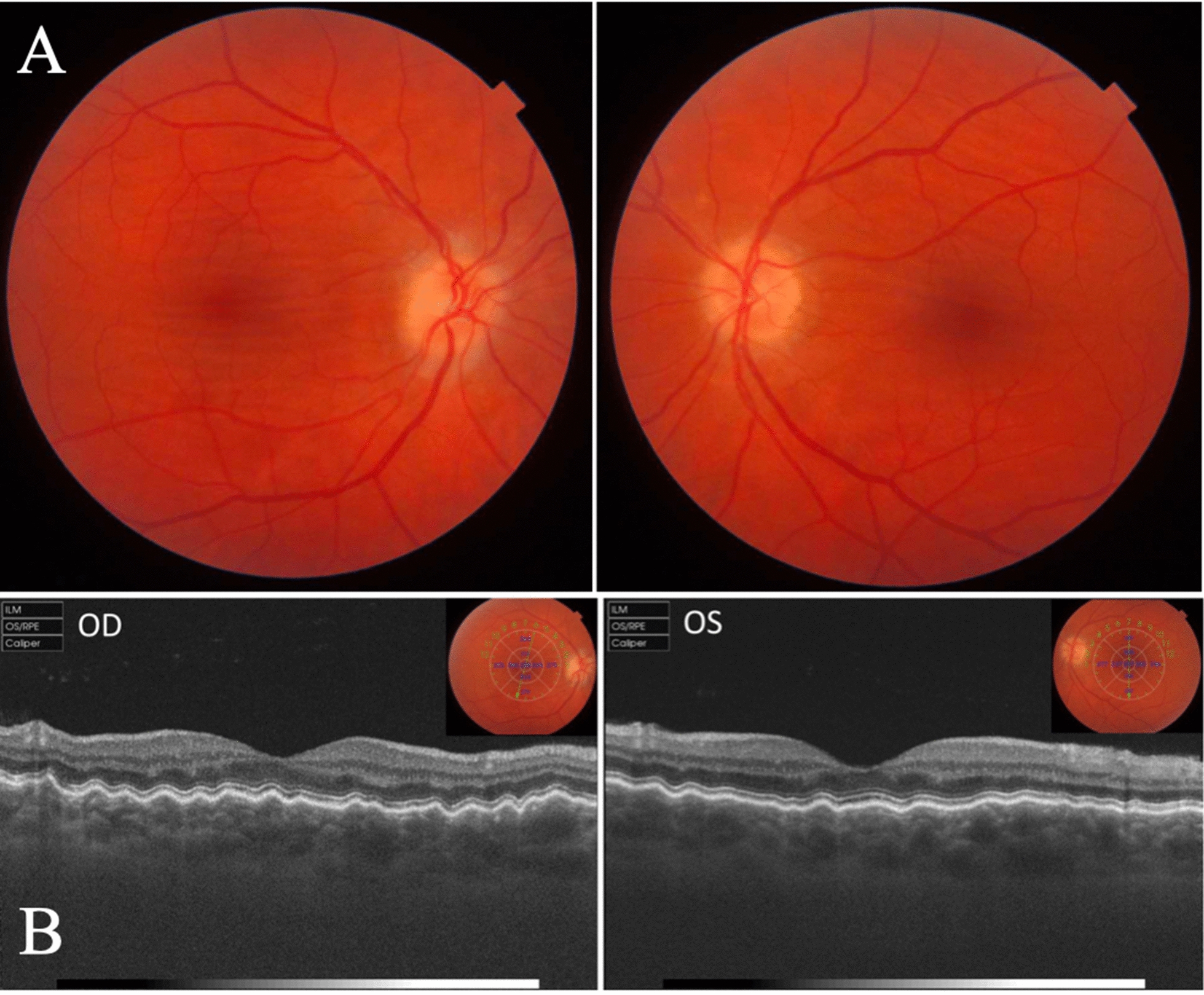
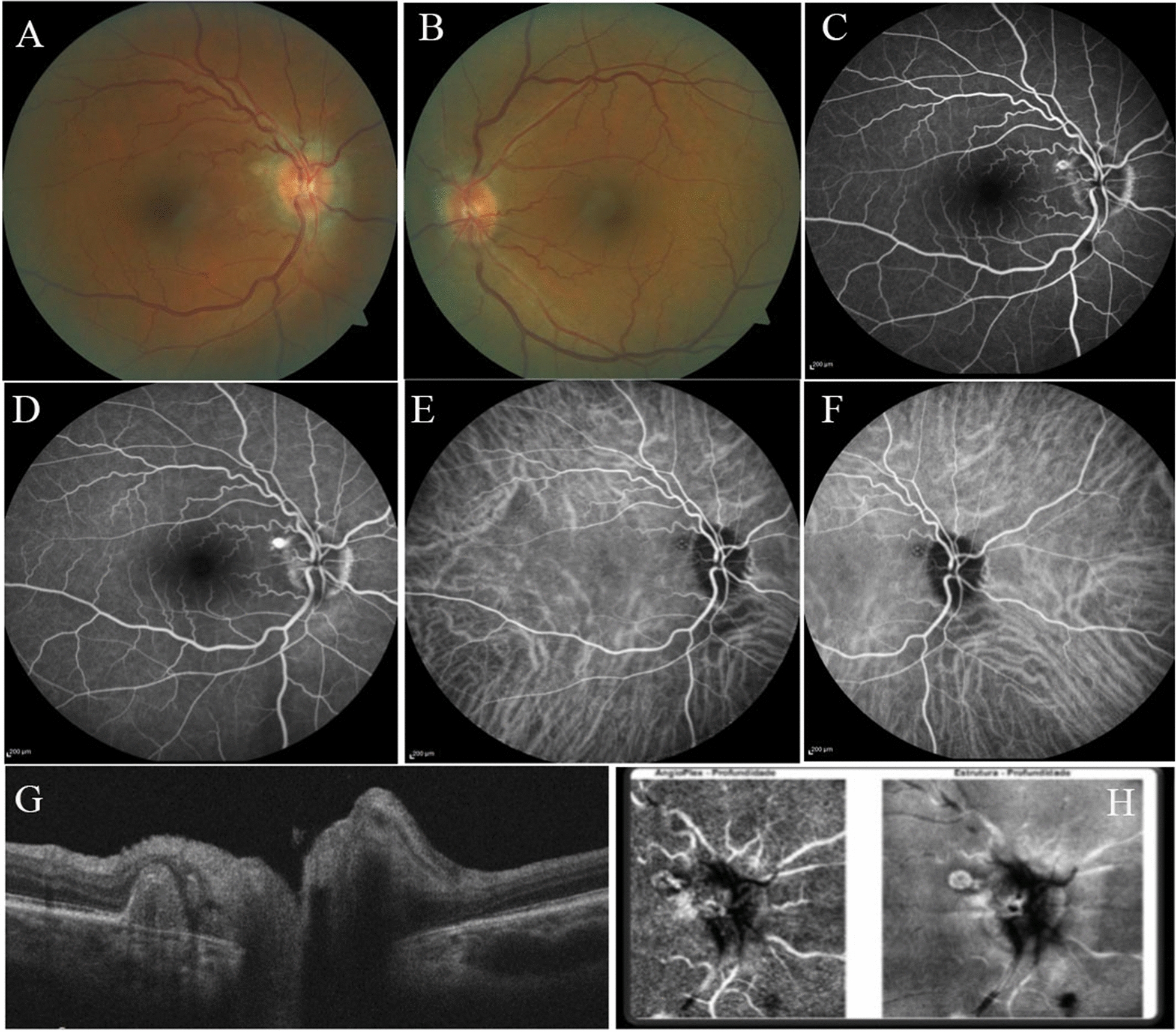
Case 1: A 47-year-old woman previous diagnosed with idiopathic intracranial hypertension treated with weight loss and acetazolamide that over the following 6 months had optic disc edema gradually resolved. The patient was follow-up for a period of 10 years and the papilledema disappeared, but choroidal folds remained unchanged. Case 2: A 61-year-old female patient was seen as a follow-up examination of a 5-year history of IIH that presented with papilledema. The patient was asymptomatic but fundoscopy evaluation revealed a yellowish white peripapillary subretinal nodular lesion temporally in OD. Multimodal imaging studies were made, and the patient was diagnosed with a rare and just recent described association of IIH and polypoidal choroidal vasculopathy.
Papilledema, RNFL and retinal ganglion cell loss are the most common structural complications of IIH, but chorioretinal complications are important findings and should be carefully evaluated in such patients. Awareness of such occurrence and the use of appropriated clinical and multimodal imaging studies are of great importance for its early detection, leading to proper treatment and prevention of further visual loss.
视乳头水肿是特发性颅内高压(IIH)患者的主要眼部表现,尽管也可能出现一些脉络膜视网膜异常并导致视力丧失。本文旨在描述两例与特发性颅内高压相关的脉络膜视网膜异常病例:一例有脉络膜皱褶,另一例有多发性脉络膜血管病变,这是该疾病中极为罕见的眼部并发症。
病例1:一名47岁女性,先前诊断为特发性颅内高压,通过减肥和乙酰唑胺治疗,在接下来的6个月中视盘水肿逐渐消退。该患者随访了10年,视乳头水肿消失,但脉络膜皱褶保持不变。病例2:一名61岁女性患者,因有5年IIH病史前来进行随访检查,表现为视乳头水肿。患者无症状,但眼底镜检查评估发现右眼颞侧视乳头周围视网膜下有一个黄白色结节状病变。进行了多模态成像研究,该患者被诊断为IIH与多发性脉络膜血管病变的一种罕见且最近才被描述的关联。
视乳头水肿、视网膜神经纤维层(RNFL)和视网膜神经节细胞丢失是IIH最常见的结构并发症,但脉络膜视网膜并发症也是重要发现,对此类患者应仔细评估。认识到这种情况的发生并使用适当的临床和多模态成像研究对于早期检测非常重要,从而能够进行适当治疗并预防进一步的视力丧失。