FIDMAG Germanes Hospitalàries Research Foundation, Barcelona, Spain; CIBERSAM (Centro de Investigación Biomédica en Red de Salud Mental), Barcelona, Spain.
Psychiatry department, Hospital Sant Rafael, Barcelona, Spain.
Neuroimage Clin. 2022;35:103119. doi: 10.1016/j.nicl.2022.103119. Epub 2022 Jul 16.
The negative symptoms of schizophrenia have been proposed to reflect prefrontal cortex dysfunction. However, this proposal has not been consistently supported in functional imaging studies, which have also used executive tasks that may not capture key aspects of negative symptoms such as lack of volition.
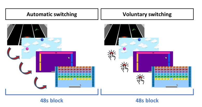
Twenty-four DSM-5 schizophrenic patients with high negative symptoms (HNS), 25 with absent negative symptoms (ANS) and 30 healthy controls underwent fMRI during performance of the Computerized Multiple Elements Test (CMET), a task designed to measure poor organization of goal directed behaviour or 'goal neglect'. Negative symptoms were rated using the PANSS and the Clinical Assessment Interview for Negative Symptoms (CAINS).
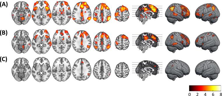
On whole brain analysis, the ANS patients showed no significant clusters of reduced activation compared to the healthy controls. In contrast, the HNS patients showed hypoactivation compared to the healthy controls in the left anterior frontal cortex, the right dorsolateral prefrontal cortex (DLPFC), the anterior insula bilaterally and the bilateral inferior parietal cortex. When compared to the ANS patients, the HNS patients showed reduced activation in the left anterior frontal cortex, the left DLPFC and the left inferior parietal cortex. After controlling for disorganization scores, differences remained in clusters in the left anterior frontal cortex and the bilateral inferior parietal cortex.
This study provides evidence that reduced prefrontal activation, perhaps especially in the left anterior frontal cortex, is a brain functional correlate of negative symptoms in schizophrenia. The simultaneous finding of reduced inferior parietal cortex activation was unexpected, but could reflect this region's involvement in cognitive control, particularly the 'regulative' component of this.
精神分裂症的阴性症状被认为反映了前额叶皮层功能障碍。然而,这一假说并未在功能影像学研究中得到一致支持,这些研究也使用了执行任务,这些任务可能无法捕捉到阴性症状的关键方面,如缺乏意志。
24 名 DSM-5 精神分裂症患者伴有高阴性症状(HNS),25 名无阴性症状(ANS)患者和 30 名健康对照者在执行计算机多元素测试(CMET)时接受 fMRI 检查,该任务旨在测量目标导向行为或“目标忽视”的组织不良。阴性症状使用 PANSS 和阴性症状临床评估访谈(CAINS)进行评估。
在全脑分析中,与健康对照组相比,ANS 患者没有明显的激活减少簇。相比之下,HNS 患者与健康对照组相比,在左侧额前皮质、右侧背外侧前额叶皮质(DLPFC)、双侧前岛叶和双侧下顶叶皮质表现出低激活。与 ANS 患者相比,HNS 患者在左侧额前皮质、左侧 DLPFC 和左侧下顶叶皮质的激活减少。在控制了组织混乱评分后,左侧额前皮质和双侧下顶叶皮质的差异仍然存在。
这项研究提供了证据,表明前额叶皮层的激活减少,尤其是左侧额前皮质,是精神分裂症阴性症状的大脑功能相关物。同时发现下顶叶皮层激活减少是出乎意料的,但可能反映了该区域在认知控制中的参与,特别是在这一成分的调节方面。