He Shanqing, Yao Yajun, Yang Nan, Wang Youcheng, Liu Dishiwen, Cao Zhen, Chen Huiyu, Fu Yuntao, Yang Mei, Wang Songjun, He Guangjie, Zhao Qingyan
Department of Cardiology, Renmin Hospital of Wuhan University, Wuhan, China.
Cardiovascular Research Institute of Wuhan University, Wuhan, China.
Front Pharmacol. 2022 Jul 7;13:925276. doi: 10.3389/fphar.2022.925276. eCollection 2022.
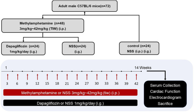
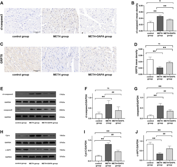
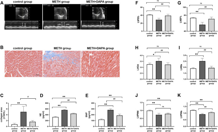
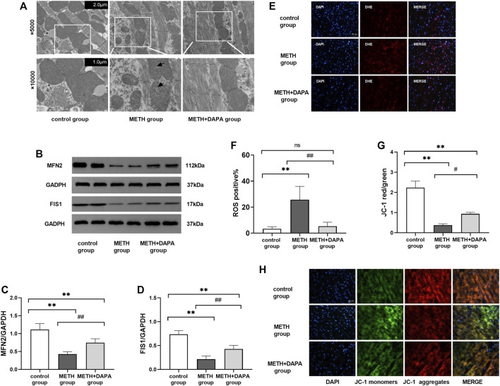
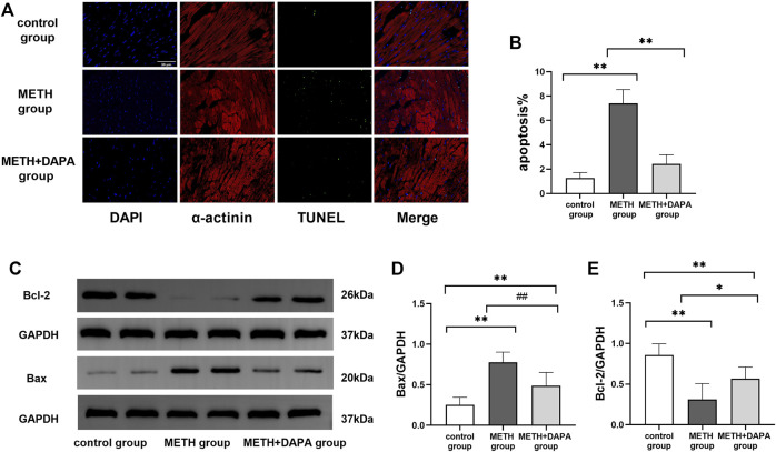
Methamphetamine (METH)-induced cardiovascular toxicity has been attributed to its destructive effect on mitochondrial function at least to some extent. Previous studies highlighted the benefits of dapagliflozin (DAPA) on the cardiovascular system, but the response of METH-induced cardiomyopathy to DAPA is never addressed before. The present study aimed to investigate the potential ability of DAPA in preventing METH-induced cardiomyopathy. C57BL/6 mice were randomly divided into control group ( = 24), METH group ( = 24), and METH + DAPA group ( = 24). The METH-induced cardiomyopathy group received intraperitoneal METH injections at gradually increasing doses thrice weekly for 14 weeks. Mice in the METH + DAPA group were simultaneously treated with DAPA 1 mg/kg/day by intragastric administration. Echocardiography was performed to assess cardiac function. Reactive oxygen species (ROS), JC-1, and terminal deoxynucleotidyl transferase dUTP nick-end labeling (TUNEL) assays were performed to evaluate oxidative stress, mitochondrial damage, and apoptosis, respectively. Mitochondrial and apoptosis-related protein expression was measured by western blotting. Mice exposed to METH exhibited reduced cardiac function (left ventricular ejection fraction [LVEF]: 56.51 ± 6.49 vs. 73.62 ± 1.42, < 0.01), fibrotic remodeling, and mitochondrial dysfunction, leading to apoptosis (apoptotic cells%: 7.4 ± 1.3 vs. 1.3 ± 0.5, < 0.01). DAPA significantly reduced mitochondrial dynamics and function, ROS, apoptosis (apoptotic cells%: 2.4 ± 0.8 vs. 7.4 ± 1.3, < 0.01), cardiac function decline (LVEF: 70.99 ± 4.936 vs. 56.51 ± 6.49, < 0.01), and fibrotic remodeling. These results indicated that DAPA could be considered as an effective therapeutic agent in the protection against METH-associated cardiomyopathy. DAPA protects against METH-induced cardiomyopathy in mice by decreasing mitochondrial damage and apoptosis.
甲基苯丙胺(METH)诱导的心血管毒性至少在一定程度上归因于其对线粒体功能的破坏作用。先前的研究强调了达格列净(DAPA)对心血管系统的益处,但METH诱导的心肌病对DAPA的反应此前从未有过相关研究。本研究旨在探讨DAPA预防METH诱导的心肌病的潜在能力。将C57BL/6小鼠随机分为对照组(n = 24)、METH组(n = 24)和METH + DAPA组(n = 24)。METH诱导的心肌病组每周三次腹腔注射逐渐增加剂量的METH,持续14周。METH + DAPA组的小鼠同时通过灌胃给予1 mg/kg/天的DAPA。进行超声心动图评估心脏功能。分别进行活性氧(ROS)、JC-1和末端脱氧核苷酸转移酶dUTP缺口末端标记(TUNEL)检测以评估氧化应激、线粒体损伤和细胞凋亡。通过蛋白质印迹法检测线粒体和凋亡相关蛋白的表达。暴露于METH的小鼠表现出心脏功能降低(左心室射血分数[LVEF]:56.51±6.49 vs. 73.62±1.42,P < 0.01)、纤维化重塑和线粒体功能障碍,导致细胞凋亡(凋亡细胞百分比:7.4±1.3 vs. 1.3±0.5,P < 0.01)。DAPA显著降低了线粒体动力学和功能、ROS、细胞凋亡(凋亡细胞百分比:2.4±0.8 vs. 7.4±1.3,P < 0.01)、心脏功能下降(LVEF:70.99±4.936 vs. 56.51±6.49,P < 0.01)以及纤维化重塑。这些结果表明,DAPA可被视为预防METH相关心肌病的有效治疗药物。DAPA通过减少线粒体损伤和细胞凋亡来保护小鼠免受METH诱导的心肌病。