Chittora Reena, Jain Ayushi, Shukla Sunil Dutt, Bhatnagar Maheep
Department of Physiology, Neurophysiology Laboratory, All India Institute of Medical Sciences, New Delhi, Delhi, India.
Department of Zoology, Animal Biotechnology and Molecular Neuroscience Laboratory, University College of Science, Mohan Lal Sukhadia University, Udaipur, Rajasthan, India.
Ann Neurosci. 2022 Jan;29(1):7-15. doi: 10.1177/09727531211059925. Epub 2022 Feb 2.
Sleep deprivation (SD) is a biological stress condition for the brain, and the pathogenesis of SD is closely related to elevated oxidative stress, mitochondrial dysfunction, a major cause of neurodegeneration. This oxidative stress-mediated cell death is attributed to rise in calcium ion influx which further excites or alters the neurotransmitters level by activating neuronal nitric oxide (NO) synthase (nNOS) release of NO in mouse SD model. This study indicates that the nitrergic neurons are possible therapeutic targets for the amelioration of SD-induced cognitive dysfunction and behavioral alterations.
SD is considered as a risk factor for various neurodegenerative diseases. SD leads to biochemical, behavioral, and neurochemical alterations in animals. This study was designed to explore the possible involvement of a nitrergic neuron system in six days SD-induced morphological and neurodegenerative changes in mice.
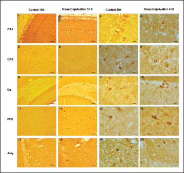
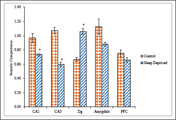
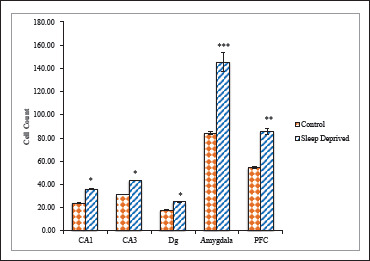
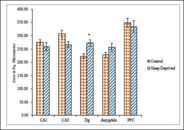
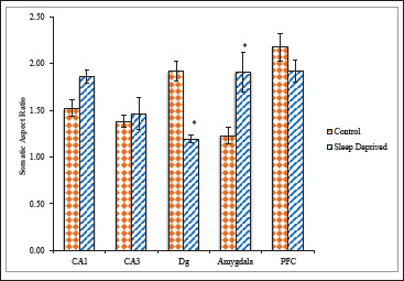
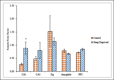
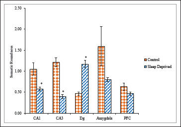
Using nNOS immunohistochemistry, we have investigated the effects of SD on nNOS positive neurons. Immunohistochemical study for the distribution of nNOS positive neuronal cell bodies was carried out in the hippocampus, prefrontal cortex (PFC), and amygdaloid nuclei of mice brain.
Sleep-deprived animals showed a significantly increased number of nNOS positive neurons and altered neuronal cytomorphology as compared with the control group.
These results indicate that total SD may induce morphological changes in nNOS positive neurons in the brain, thus increasing NO synthesis, which is implicated in SD-induced neuronal cell death.
睡眠剥夺(SD)是大脑的一种生物应激状态,SD的发病机制与氧化应激升高、线粒体功能障碍密切相关,而线粒体功能障碍是神经退行性变的主要原因。这种氧化应激介导的细胞死亡归因于钙离子内流增加,钙离子内流通过激活神经元型一氧化氮合酶(nNOS)释放一氧化氮(NO),进一步兴奋或改变神经递质水平,这在小鼠SD模型中得到证实。本研究表明,含氮能神经元可能是改善SD诱导的认知功能障碍和行为改变的治疗靶点。
SD被认为是各种神经退行性疾病的一个危险因素。SD会导致动物出现生化、行为和神经化学改变。本研究旨在探讨含氮能神经元系统在六天SD诱导的小鼠形态学和神经退行性变中的可能作用。
我们采用nNOS免疫组织化学方法,研究了SD对nNOS阳性神经元的影响。对小鼠脑内海马体、前额叶皮质(PFC)和杏仁核中nNOS阳性神经元细胞体的分布进行了免疫组织化学研究。
与对照组相比,睡眠剥夺动物的nNOS阳性神经元数量显著增加,神经元细胞形态发生改变。
这些结果表明,完全睡眠剥夺可能诱导大脑中nNOS阳性神经元的形态学改变,从而增加NO的合成,这与SD诱导的神经元细胞死亡有关。