Westlake Laboratory of Life Sciences and Biomedicine, Key Laboratory of Structural Biology of Zhejiang Province, School of Life Sciences, Westlake University, Hangzhou 310024, China.
Institute of Biology, Westlake Institute for Advanced Study, Hangzhou 310024, China.
Proc Natl Acad Sci U S A. 2022 Aug 16;119(33):e2209164119. doi: 10.1073/pnas.2209164119. Epub 2022 Jul 25.
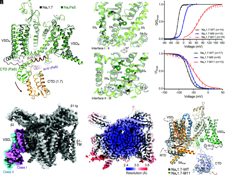
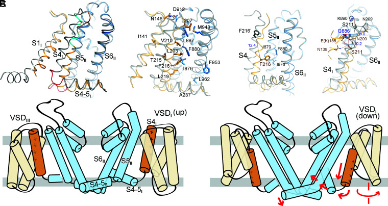
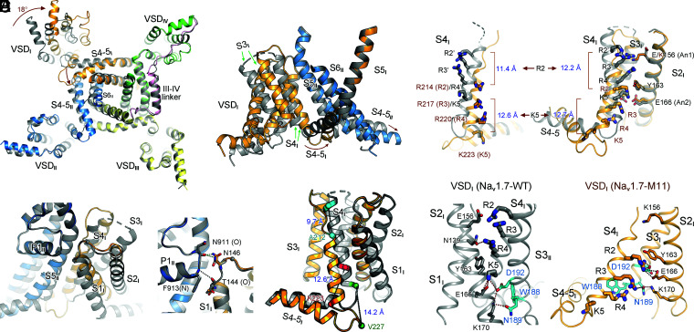
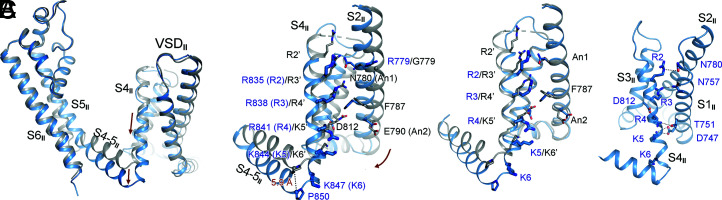
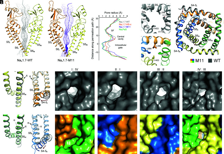
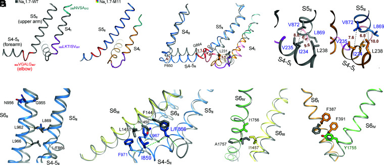
Voltage-gated sodium (Na) channel Na1.7 has been targeted for the development of nonaddictive pain killers. Structures of Na1.7 in distinct functional states will offer an advanced mechanistic understanding and aid drug discovery. Here we report the cryoelectron microscopy analysis of a human Na1.7 variant that, with 11 rationally introduced point mutations, has a markedly right-shifted activation voltage curve with V reaching 69 mV. The voltage-sensing domain in the first repeat (VSD) in a 2.7-Å resolution structure displays a completely down (deactivated) conformation. Compared to the structure of WT Na1.7, three gating charge (GC) residues in VSD are transferred to the cytosolic side through a combination of helix unwinding and spiral sliding of S4 and ∼20° domain rotation. A conserved WNФФD motif on the cytoplasmic end of S3 stabilizes the down conformation of VSD. One GC residue is transferred in VSD mainly through helix sliding. Accompanying GC transfer in VSD and VSD, rearrangement and contraction of the intracellular gate is achieved through concerted movements of adjacent segments, including S4-5, S4-5, S5, and all S6 segments. Our studies provide important insight into the electromechanical coupling mechanism of the single-chain voltage-gated ion channels and afford molecular interpretations for a number of pain-associated mutations whose pathogenic mechanism cannot be revealed from previously reported Na structures.
电压门控钠离子(Na)通道 Na1.7 已成为开发非成瘾性止痛药的靶点。处于不同功能状态的 Na1.7 的结构将提供更深入的机械理解,并有助于药物发现。在这里,我们报告了一种人源 Na1.7 变体的冷冻电镜分析,该变体通过 11 个合理引入的点突变,具有明显向右移动的激活电压曲线,V 达到 69 mV。在 2.7-Å 分辨率结构中,第一个重复(VSD)的电压感应结构域显示出完全向下(失活)的构象。与 WT Na1.7 的结构相比,VSD 中的三个门控电荷(GC)残基通过 S4 和 ∼20°结构域旋转的螺旋滑动和螺旋展开组合,转移到胞质侧。S3 胞质末端的保守 WNФФD 基序稳定 VSD 的向下构象。一个 GC 残基主要通过螺旋滑动转移到 VSD 中。伴随着 VSD 和 VSD 中的 GC 转移,通过相邻片段的协调运动,包括 S4-5、S4-5、S5 和所有 S6 片段,实现了细胞内门的重排和收缩。我们的研究为单链电压门控离子通道的机电耦联机制提供了重要的见解,并为许多疼痛相关突变提供了分子解释,这些突变的发病机制无法从以前报道的 Na 结构中揭示。