Graduate Program in Dentistry, Federal University of Pelotas, Rua Gonçalves Chaves 457, Pelotas, RS, 96015-560, Brazil.
Radboud Institute for Health Sciences, Department of Dentistry, Radboud University Medical Center, Ph. van Leydenlaan 25, NL 6500 HB, P.O. Box 9101, Nijmegen, 6525 EX, The Netherlands.
Clin Oral Investig. 2022 Dec;26(12):6925-6939. doi: 10.1007/s00784-022-04647-y. Epub 2022 Jul 26.
Deterioration in anterior resin composite restorations placed in tooth wear patients was investigated after 36 months.
Data collected prospectively for 47 participants of the Radboud Tooth Wear Project were used (41 ± 8 years, 90% male, n = 270 restorations). Restorations were individually evaluated using intraoral photographs and 3D scans to rate modified FDI scores and to record the presence of degradation features. Four groups with distinct combinations of composites and techniques were assessed, and multivariable logistic regression models were used to analyze the data (p < 0.05).
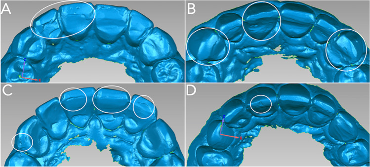
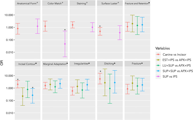
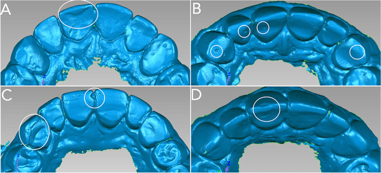
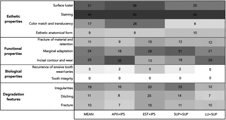
For all groups together, early degradation signs were present at 1 month: irregularities (41.5%) and ditching (7.4%) were observed at the surface and adhesive interfaces. The frequency of irregularities decreased in the 36-month evaluation (37%), but ditching (12.2%) and fractures (10.7%) were more common. The most frequent deterioration (based on photographs) was observed for staining (44%) and loss of luster (31%). In 3D scans, the most frequent were for wear (25%), marginal adaptation (24%), and the presence of irregularities (19%). Canines had 5.5 times more chances of deterioration by ditching than incisors (p < 0.001). The differences between composites and restorative techniques were minor.
A continuous degradation process of restorations placed in tooth wear patients was observed in anterior teeth restored with different composites, with a progression of the deterioration over 36 months.
When placing anterior resin composite restorations in tooth wear patients, it could be important to establish realistic expectations and the need for checkup appointments.
研究牙磨损患者的前牙树脂复合修复体在 36 个月后的恶化情况。
使用前瞻性收集的 Radboud 牙磨损项目 47 名参与者的数据(41±8 岁,90%为男性,n=270 个修复体)。使用口腔内照片和 3D 扫描对修复体进行单独评估,以评估改良 FDI 评分,并记录降解特征的存在。评估了四个具有不同复合材料和技术组合的组,并使用多变量逻辑回归模型分析数据(p<0.05)。
对于所有组,在 1 个月时就出现了早期降解迹象:表面和黏接界面出现不平整(41.5%)和刻痕(7.4%)。在 36 个月的评估中,不平整的频率降低(37%),但刻痕(12.2%)和裂缝(10.7%)更为常见。基于照片,最常见的恶化是染色(44%)和失光(31%)。在 3D 扫描中,最常见的是磨损(25%)、边缘适应性(24%)和不平整(19%)。与切牙相比,犬齿因刻痕而恶化的几率高 5.5 倍(p<0.001)。复合材料和修复技术之间的差异较小。
在牙磨损患者的前牙中使用不同的复合材料进行修复后,观察到修复体的持续降解过程,在 36 个月的时间里,恶化情况逐渐加重。
在牙磨损患者中放置前牙树脂复合修复体时,建立现实的期望和定期检查的需求可能很重要。