Department of Systems Biology, University of Alcala, 28801 Madrid, Spain.
Visual Neurophysiology Group-IRYCIS, 28034 Madrid, Spain.
Int J Mol Sci. 2022 Jul 24;23(15):8152. doi: 10.3390/ijms23158152.
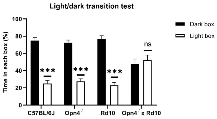
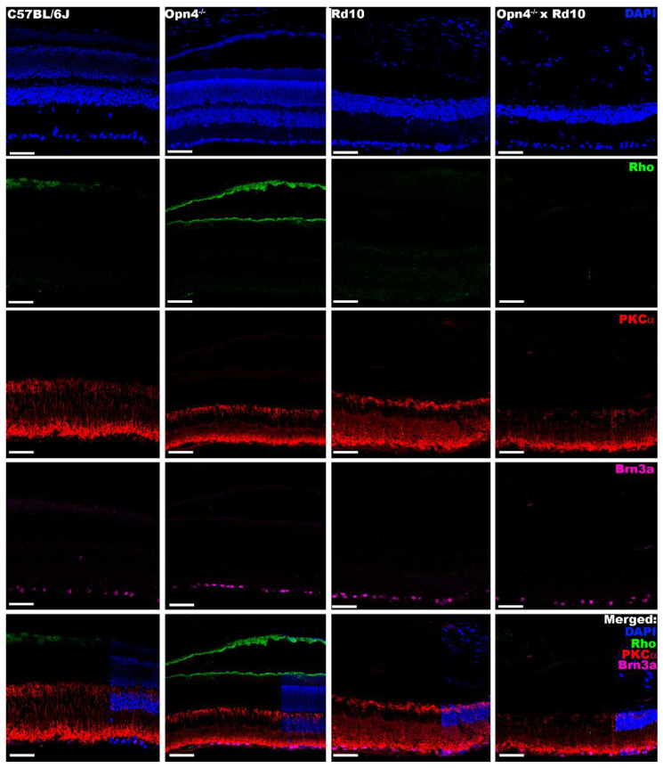
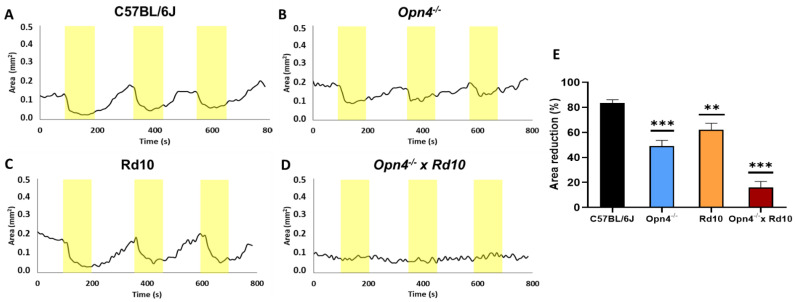
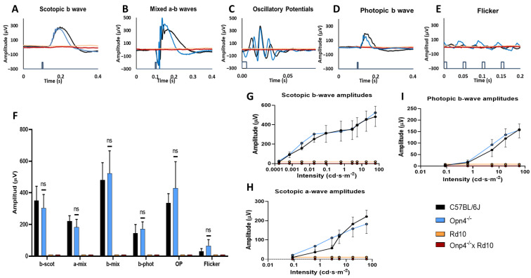
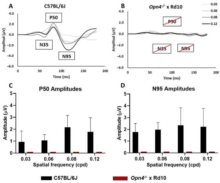
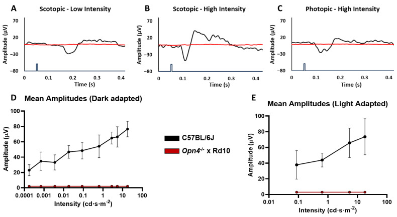
Recent technological development requires new approaches to address the problem of blindness. Such approaches need to be able to ensure that no cells with photosensitive capability remain in the retina. The presented model, × (O×Rd) double mutant murine, is a combination of a mutation in the gene (photoreceptor degeneration) together with a deletion of the gene (responsible for the expression of melanopsin in the intrinsically photosensitive retinal ganglion cells). This model has been characterized and compared with those of WT mice and murine animal models displaying both mutations separately. A total loss of pupillary reflex was observed. Likewise, behavioral tests demonstrated loss of rejection to illuminated spaces and a complete decrease in visual acuity (optomotor test). Functional recordings showed an absolute disappearance of various wave components of the full-field and pattern electroretinogram (fERG, pERG). Likewise, visual evoked potential (VEP) could not be recorded. Immunohistochemical staining showed marked degeneration of the outer retinal layers and the absence of melanopsin staining. The combination of both mutations has generated an animal model that does not show any photosensitive element in its retina. This model is a potential tool for the study of new ophthalmological approaches such as optosensitive agents.
最近的技术发展需要新的方法来解决失明问题。这些方法需要能够确保视网膜中没有感光能力的细胞残留。所提出的模型, × (O×Rd)双突变鼠,是一个突变的组合在 基因(感光细胞变性)与 基因缺失(负责内在感光视网膜神经节细胞中黑视素的表达)。该模型已被表征,并与 WT 小鼠和单独显示两种突变的鼠动物模型进行了比较。观察到瞳孔反射完全丧失。同样,行为测试表明对光照空间的排斥反应丧失,以及视力(光动测试)完全下降。功能记录显示全视野和图形视网膜电图(fERG,pERG)的各种波成分绝对消失。同样,无法记录视觉诱发电位(VEP)。免疫组织化学染色显示外视网膜层明显变性,并且黑视素染色缺失。两种突变的组合产生了一种在其视网膜中没有任何感光元件的动物模型。该模型是研究新的眼科方法(如光敏剂)的潜在工具。