• 文献检索
  • 文档翻译
  • 深度研究
  • 学术资讯
  • Suppr Zotero 插件Zotero 插件
  • 邀请有礼
  • 套餐&价格
  • 历史记录
应用&插件
Suppr Zotero 插件Zotero 插件浏览器插件Mac 客户端Windows 客户端微信小程序
定价
高级版会员购买积分包购买API积分包
服务
文献检索文档翻译深度研究API 文档MCP 服务
关于我们
关于 Suppr公司介绍联系我们用户协议隐私条款
关注我们

Suppr 超能文献

核心技术专利:CN118964589B侵权必究
粤ICP备2023148730 号-1Suppr @ 2026

文献检索

告别复杂PubMed语法,用中文像聊天一样搜索,搜遍4000万医学文献。AI智能推荐,让科研检索更轻松。

立即免费搜索

文件翻译

保留排版,准确专业,支持PDF/Word/PPT等文件格式,支持 12+语言互译。

免费翻译文档

深度研究

AI帮你快速写综述,25分钟生成高质量综述,智能提取关键信息,辅助科研写作。

立即免费体验

pSK1 质粒的分子分析:一种新型由葡萄球菌多重耐药质粒编码的质粒分配系统。

Molecular Analysis of pSK1 par: A Novel Plasmid Partitioning System Encoded by Staphylococcal Multiresistance Plasmids.

机构信息

School of Life and Environmental Sciences, University of Sydney, New South Wales 2006, Australia.

Microbiology and Infectious Diseases, School of Medicine, Western Sydney University, New South Wales 2751, Australia; Antibiotic Resistance & Mobile Elements Group, Ingham Institute for Applied Medical Research, New South Wales 2170, Australia.

出版信息

J Mol Biol. 2022 Oct 15;434(19):167770. doi: 10.1016/j.jmb.2022.167770. Epub 2022 Jul 27.

DOI:10.1016/j.jmb.2022.167770
PMID:35907571
原文链接:https://pmc.ncbi.nlm.nih.gov/articles/PMC9595883/
Abstract

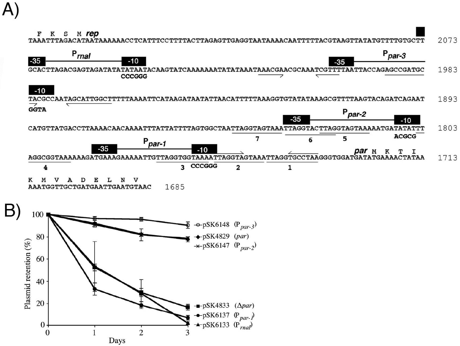
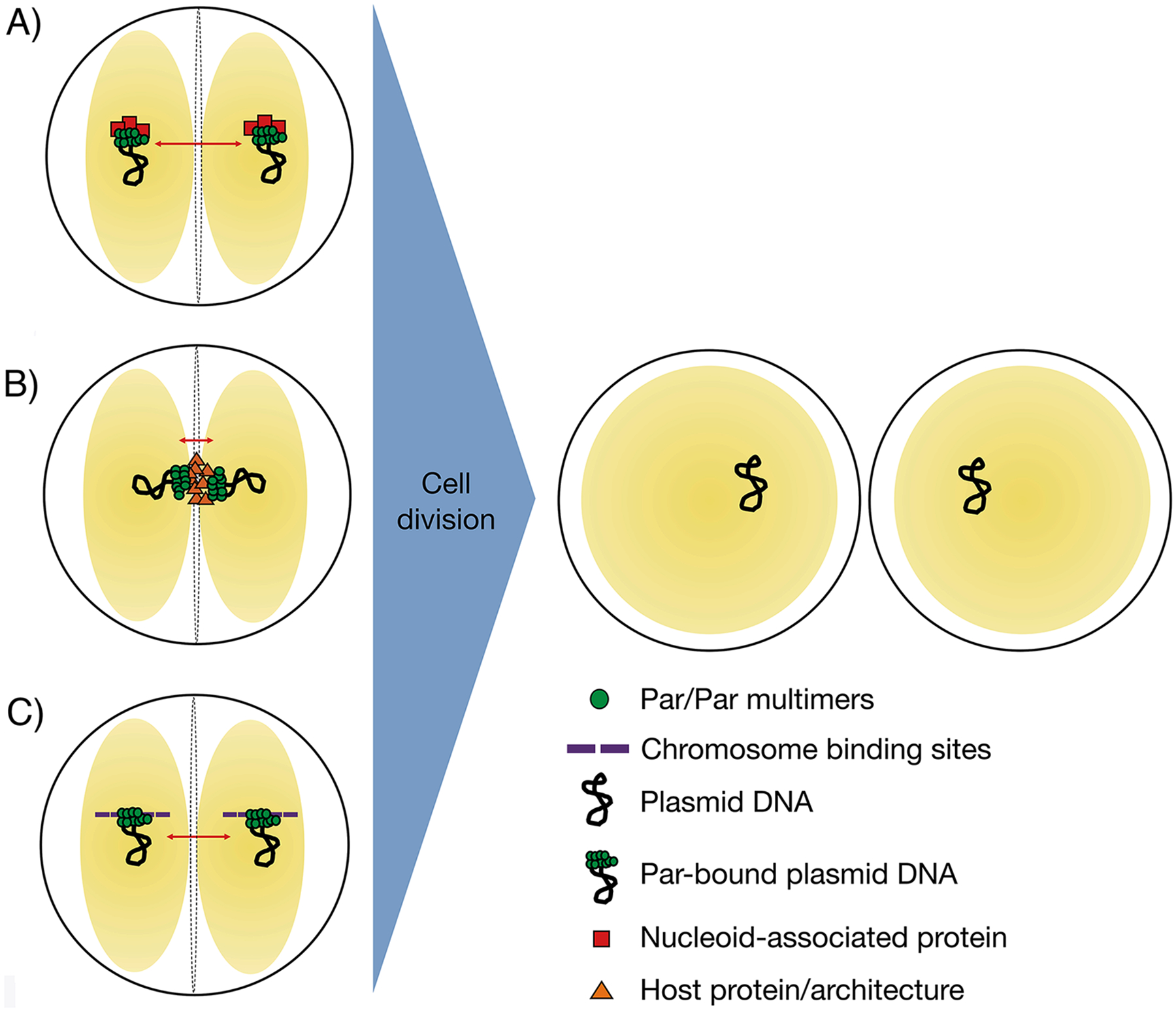
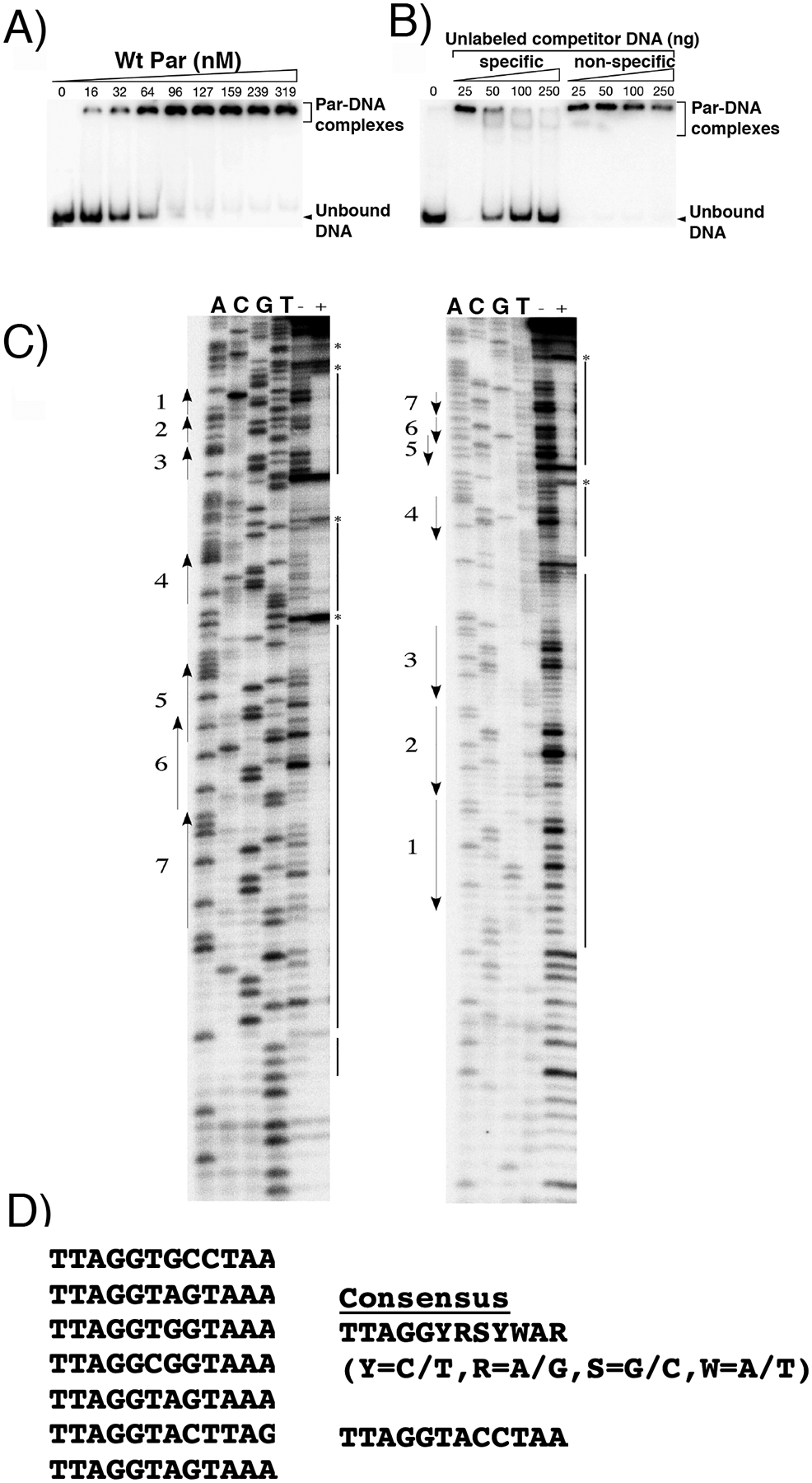
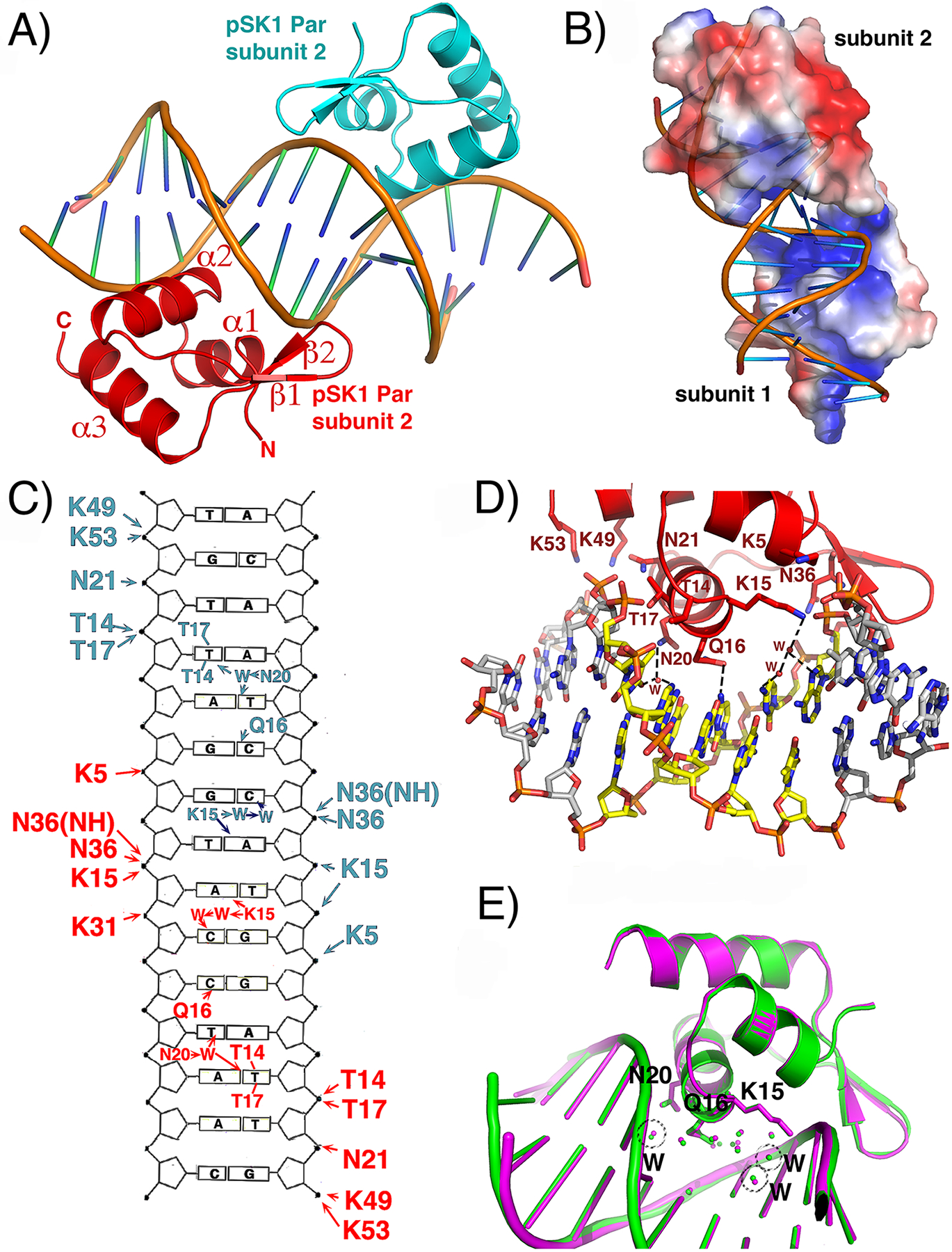
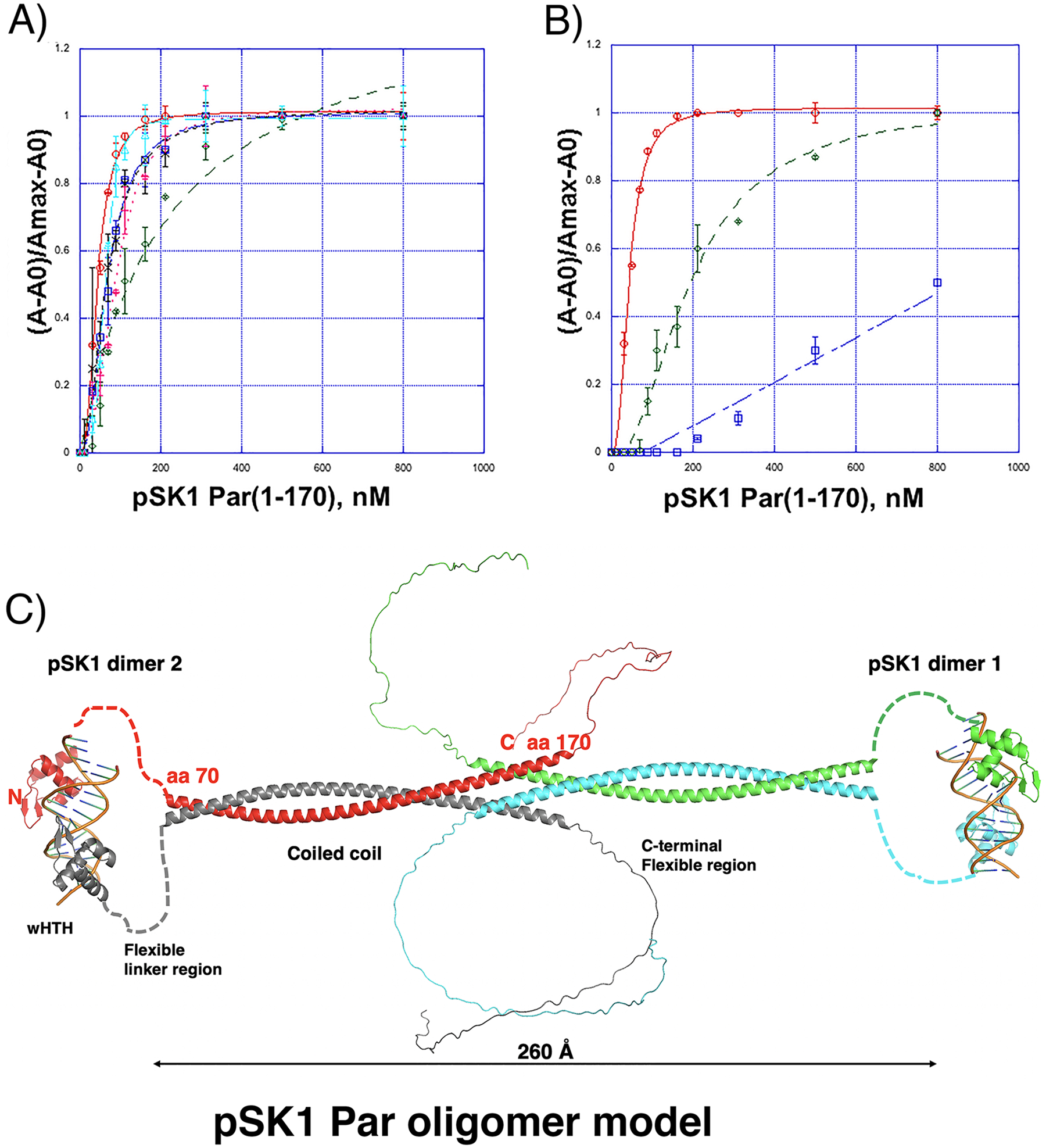
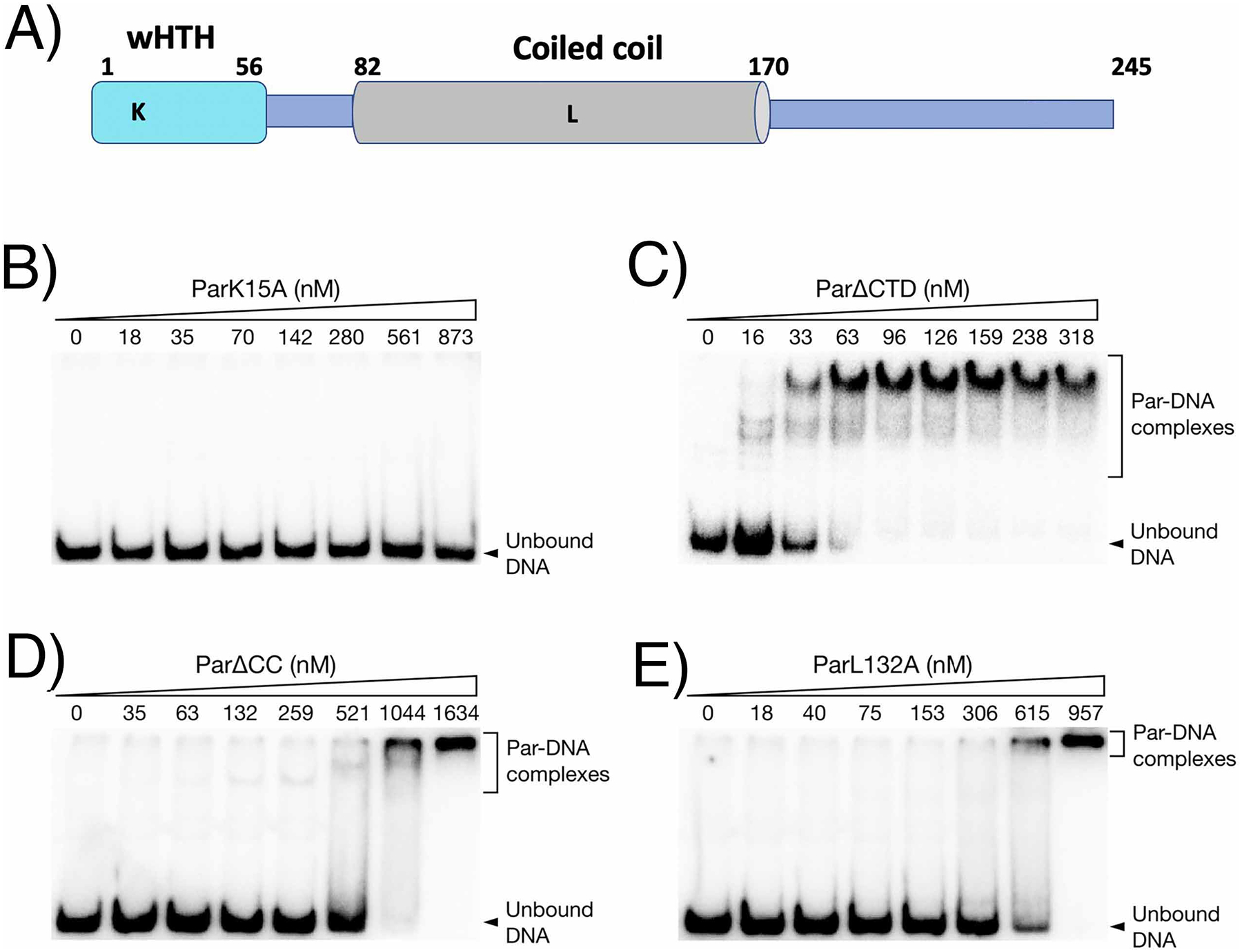
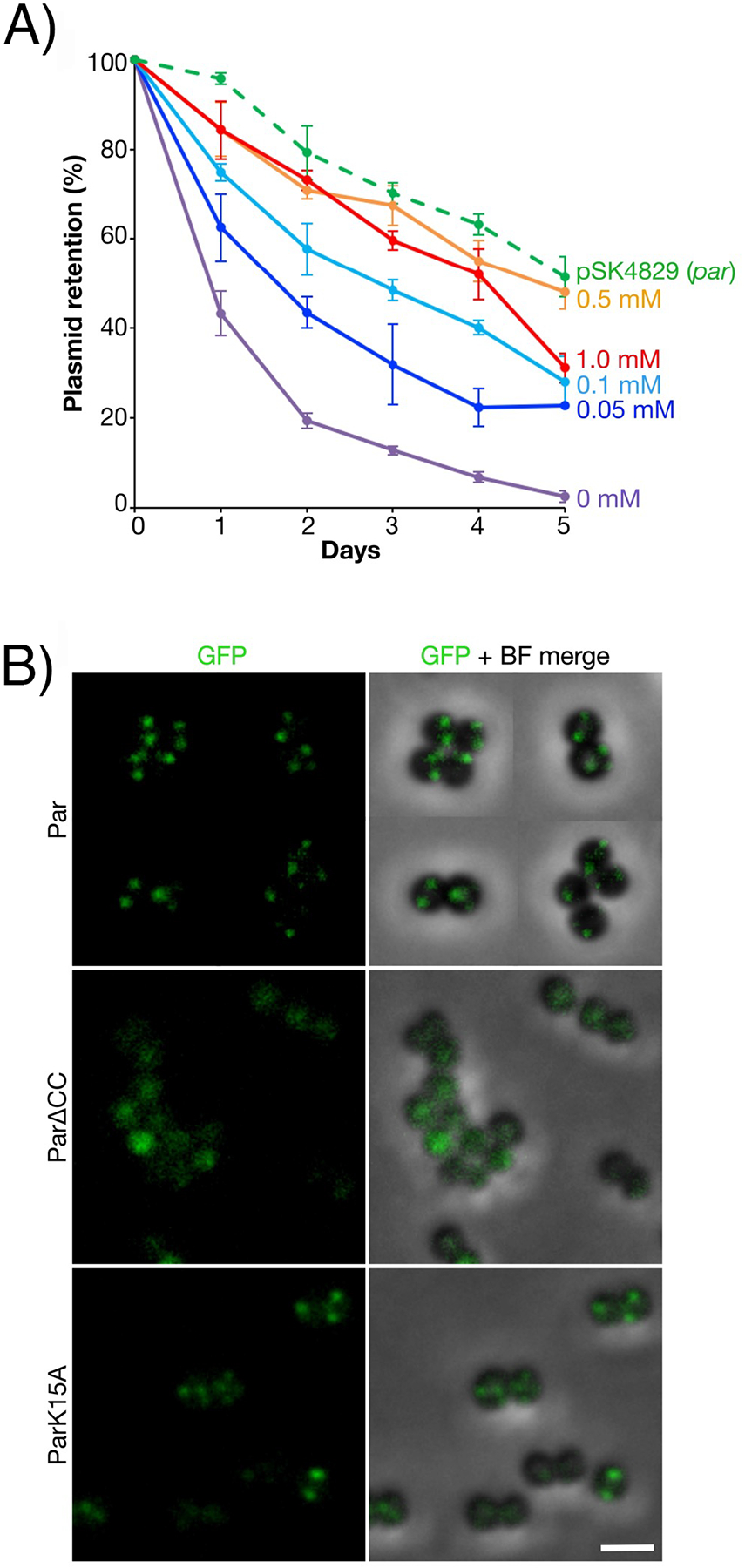
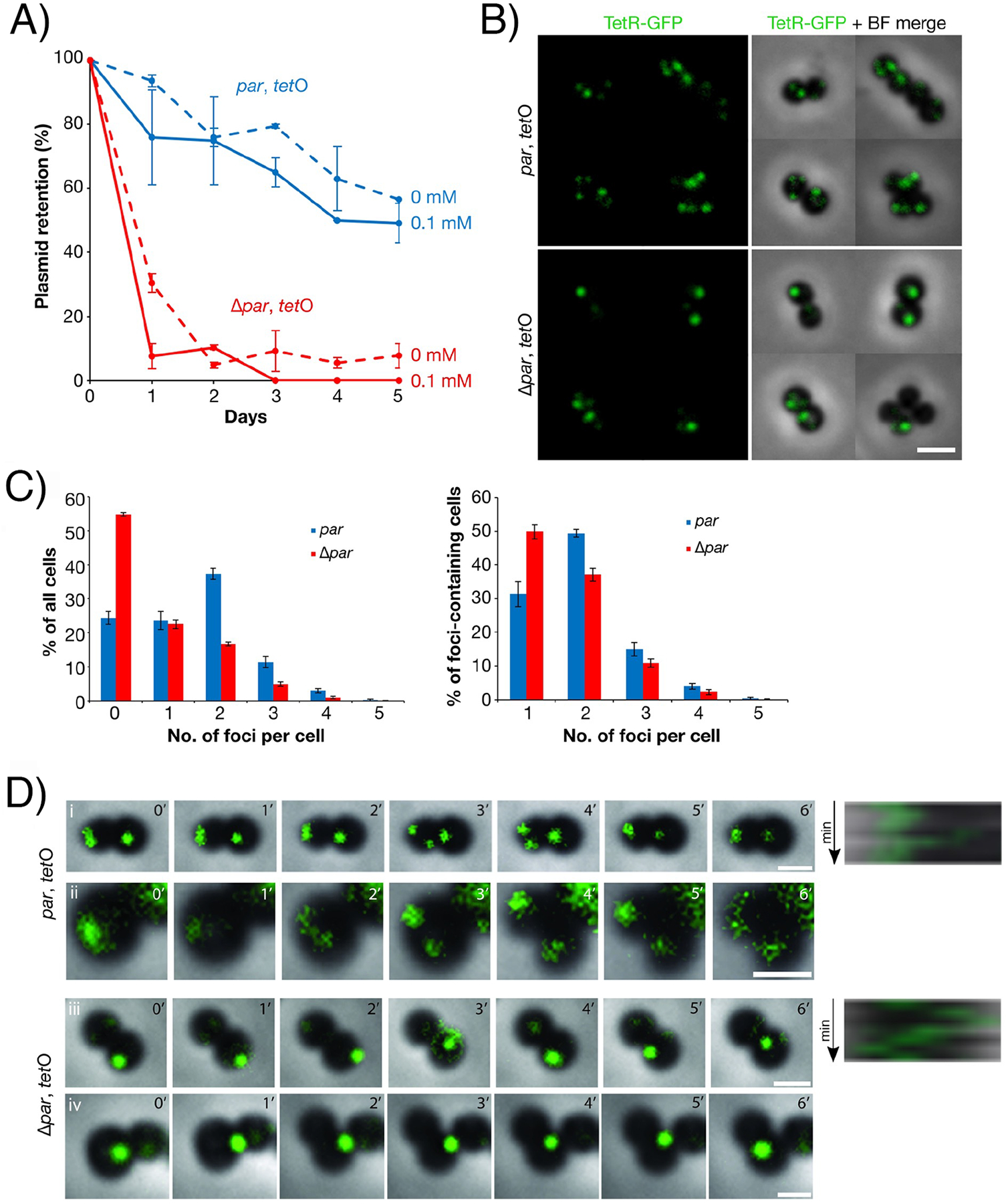
The segregation of prokaryotic plasmids typically requires a centromere-like site and two proteins, a centromere-binding protein (CBP) and an NTPase. By contrast, a single 245 residue Par protein mediates partition of the prototypical staphylococcal multiresistance plasmid pSK1 in the absence of an identifiable NTPase component. To gain insight into centromere binding by pSK1 Par and its segregation function we performed structural, biochemical and in vivo studies. Here we show that pSK1 Par binds a centromere consisting of seven repeat elements. We demonstrate this Par-centromere interaction also mediates Par autoregulation. To elucidate the Par centromere binding mechanism, we obtained a structure of the Par N-terminal DNA-binding domain bound to centromere DNA to 2.25 Å. The pSK1 Par structure, which harbors a winged-helix-turn-helix (wHTH), is distinct from other plasmid CBP structures but shows homology to the B. subtilis chromosome segregation protein, RacA. Biochemical studies suggest the region C-terminal to the Par wHTH forms coiled coils and mediates oligomerization. Fluorescence microscopy analyses show that pSK1 Par enhances the separation of plasmids from clusters, driving effective segregation upon cell division. Combined the data provide insight into the molecular properties of a single protein partition system.

摘要

原核质体的分离通常需要一个类似于着丝粒的位点和两种蛋白质,即着丝粒结合蛋白(CBP)和 NTP 酶。相比之下,单个 245 个残基的 Par 蛋白在没有可识别的 NTP 酶成分的情况下,介导了典型的葡萄球菌多耐药质体 pSK1 的分配。为了深入了解 pSK1 Par 对着丝粒的结合及其分离功能,我们进行了结构、生化和体内研究。在这里,我们显示 pSK1 Par 结合由七个重复元件组成的着丝粒。我们证明这种 Par-着丝粒相互作用也介导 Par 自身调节。为了阐明 Par 着丝粒结合机制,我们获得了 Par N 端 DNA 结合结构域与 2.25Å 的着丝粒 DNA 结合的结构。pSK1 Par 结构含有一个翼状螺旋-转角-螺旋(wHTH),与其他质体 CBP 结构不同,但与 B. subtilis 染色体分离蛋白 RacA 具有同源性。生化研究表明,Par wHTH 结构域 C 端形成卷曲螺旋,并介导寡聚化。荧光显微镜分析表明,pSK1 Par 增强了质粒与簇的分离,在细胞分裂时促进有效分离。综合这些数据,深入了解了单个蛋白分配系统的分子特性。

https://cdn.ncbi.nlm.nih.gov/pmc/blobs/a650/9595883/62e4a2dbe527/nihms-1842025-f0008.jpg
https://cdn.ncbi.nlm.nih.gov/pmc/blobs/a650/9595883/027ffb570af2/nihms-1842025-f0001.jpg
https://cdn.ncbi.nlm.nih.gov/pmc/blobs/a650/9595883/4bf54e99114f/nihms-1842025-f0002.jpg
https://cdn.ncbi.nlm.nih.gov/pmc/blobs/a650/9595883/ac1bcb5e61a3/nihms-1842025-f0003.jpg
https://cdn.ncbi.nlm.nih.gov/pmc/blobs/a650/9595883/eec1a381db9b/nihms-1842025-f0004.jpg
https://cdn.ncbi.nlm.nih.gov/pmc/blobs/a650/9595883/32c4ce5b0bda/nihms-1842025-f0005.jpg
https://cdn.ncbi.nlm.nih.gov/pmc/blobs/a650/9595883/fd53302eb931/nihms-1842025-f0006.jpg
https://cdn.ncbi.nlm.nih.gov/pmc/blobs/a650/9595883/7ed2f733df37/nihms-1842025-f0007.jpg
https://cdn.ncbi.nlm.nih.gov/pmc/blobs/a650/9595883/62e4a2dbe527/nihms-1842025-f0008.jpg

相似文献

1
Molecular Analysis of pSK1 par: A Novel Plasmid Partitioning System Encoded by Staphylococcal Multiresistance Plasmids.pSK1 质粒的分子分析:一种新型由葡萄球菌多重耐药质粒编码的质粒分配系统。
J Mol Biol. 2022 Oct 15;434(19):167770. doi: 10.1016/j.jmb.2022.167770. Epub 2022 Jul 27.
2
Atypical low-copy number plasmid segregation systems, all in one?非典型低拷贝数质粒分离系统,集于一体?
Plasmid. 2023 Jul;127:102694. doi: 10.1016/j.plasmid.2023.102694. Epub 2023 Jun 8.
3
A single gene on the staphylococcal multiresistance plasmid pSK1 encodes a novel partitioning system.葡萄球菌多重耐药质粒pSK1上的一个单一基因编码一种新型的分配系统。
J Bacteriol. 2003 Apr;185(7):2143-52. doi: 10.1128/JB.185.7.2143-2152.2003.
4
Structural biology of plasmid partition: uncovering the molecular mechanisms of DNA segregation.质粒分配的结构生物学:揭示DNA分离的分子机制
Biochem J. 2008 May 15;412(1):1-18. doi: 10.1042/BJ20080359.
5
Plasmid protein TubR uses a distinct mode of HTH-DNA binding and recruits the prokaryotic tubulin homolog TubZ to effect DNA partition.质粒蛋白 TubR 使用独特的 HTH-DNA 结合模式招募原核微管蛋白同源物 TubZ 以实现 DNA 分配。
Proc Natl Acad Sci U S A. 2010 Jun 29;107(26):11763-8. doi: 10.1073/pnas.1003817107. Epub 2010 Jun 4.
6
Segrosome structure revealed by a complex of ParR with centromere DNA.ParR与着丝粒DNA复合物揭示的分割体结构。
Nature. 2007 Dec 20;450(7173):1268-71. doi: 10.1038/nature06392.
7
The C-terminal region of the plasmid partitioning protein TubY is a tetramer that can bind membranes and DNA.质粒分配蛋白 TubY 的 C 端区域是一个可以结合膜和 DNA 的四聚体。
J Biol Chem. 2020 Dec 18;295(51):17770-17780. doi: 10.1074/jbc.RA120.014705.
8
Characterization of the DNA Binding Domain of StbA, A Key Protein of A New Type of DNA Segregation System.StbA 的 DNA 结合结构域的特性研究,StbA 是一种新型 DNA 分离系统的关键蛋白。
J Mol Biol. 2022 Oct 15;434(19):167752. doi: 10.1016/j.jmb.2022.167752. Epub 2022 Jul 19.
9
Centromere binding and evolution of chromosomal partition systems in the Burkholderiales.博克霍尔德氏菌目的着丝粒结合和染色体分区系统的演化。
J Bacteriol. 2012 Jul;194(13):3426-36. doi: 10.1128/JB.00041-12. Epub 2012 Apr 20.
10
Structural biology of plasmid segregation proteins.质粒分离蛋白的结构生物学
Curr Opin Struct Biol. 2007 Feb;17(1):103-9. doi: 10.1016/j.sbi.2006.11.005. Epub 2006 Dec 11.

引用本文的文献

1
Molecular dissection of the chromosome partitioning protein RocS and regulation by phosphorylation.染色体分配蛋白 RocS 的分子剖析及磷酸化调控。
J Bacteriol. 2024 Oct 24;206(10):e0029124. doi: 10.1128/jb.00291-24. Epub 2024 Sep 24.
2
DNA Segregation in Enterobacteria.肠杆菌中的DNA分离
EcoSal Plus. 2023 Dec 12;11(1):eesp00382020. doi: 10.1128/ecosalplus.esp-0038-2020. Epub 2023 May 9.

本文引用的文献

1
ColabFold: making protein folding accessible to all.ColabFold:让蛋白质折叠变得人人可用。
Nat Methods. 2022 Jun;19(6):679-682. doi: 10.1038/s41592-022-01488-1. Epub 2022 May 30.
2
AlphaFold Protein Structure Database: massively expanding the structural coverage of protein-sequence space with high-accuracy models.AlphaFold 蛋白质结构数据库:用高精度模型极大地扩展蛋白质序列空间的结构覆盖范围。
Nucleic Acids Res. 2022 Jan 7;50(D1):D439-D444. doi: 10.1093/nar/gkab1061.
3
Relief of ParB autoinhibition by DNA catalysis and recycling of ParB by CTP hydrolysis promote bacterial centromere assembly.
通过DNA催化解除ParB自身抑制以及通过CTP水解实现ParB循环利用,促进细菌着丝粒组装。
Sci Adv. 2021 Oct 8;7(41):eabj2854. doi: 10.1126/sciadv.abj2854. Epub 2021 Oct 6.
4
The CTPase activity of ParB determines the size and dynamics of prokaryotic DNA partition complexes.ParB 的 CTPase 活性决定了原核 DNA 分区复合物的大小和动态。
Mol Cell. 2021 Oct 7;81(19):3992-4007.e10. doi: 10.1016/j.molcel.2021.09.004. Epub 2021 Sep 24.
5
A CTP-dependent gating mechanism enables ParB spreading on DNA.CTP 依赖性门控机制使 ParB 在 DNA 上扩散。
Elife. 2021 Aug 16;10:e69676. doi: 10.7554/eLife.69676.
6
CTP and coordinate ParB partition complex dynamics and ParA-ATPase activation for ParABS-mediated DNA partitioning.CTP 与 ParB 分隔复合动力学协同作用,激活 ParA-ATP 酶,以实现 ParABS 介导的 DNA 分隔。
Elife. 2021 Jul 21;10:e65651. doi: 10.7554/eLife.65651.
7
Highly accurate protein structure prediction with AlphaFold.利用 AlphaFold 进行高精度蛋白质结构预测。
Nature. 2021 Aug;596(7873):583-589. doi: 10.1038/s41586-021-03819-2. Epub 2021 Jul 15.
8
ParB-type DNA Segregation Proteins Are CTP-Dependent Molecular Switches.ParB 型 DNA 分离蛋白是依赖于 CTP 的分子开关。
Cell. 2019 Dec 12;179(7):1512-1524.e15. doi: 10.1016/j.cell.2019.11.015.
9
Self-organization of centromeres by the ParB CTP hydrolase.着丝粒的 ParB CTP 水解酶的自组织。
Science. 2019 Nov 29;366(6469):1129-1133. doi: 10.1126/science.aay3965. Epub 2019 Oct 24.
10
Web 3DNA 2.0 for the analysis, visualization, and modeling of 3D nucleic acid structures.Web 3DNA 2.0 用于分析、可视化和建模 3D 核酸结构。
Nucleic Acids Res. 2019 Jul 2;47(W1):W26-W34. doi: 10.1093/nar/gkz394.