• 文献检索
  • 文档翻译
  • 深度研究
  • 学术资讯
  • Suppr Zotero 插件Zotero 插件
  • 邀请有礼
  • 套餐&价格
  • 历史记录
应用&插件
Suppr Zotero 插件Zotero 插件浏览器插件Mac 客户端Windows 客户端微信小程序
定价
高级版会员购买积分包购买API积分包
服务
文献检索文档翻译深度研究API 文档MCP 服务
关于我们
关于 Suppr公司介绍联系我们用户协议隐私条款
关注我们

Suppr 超能文献

核心技术专利:CN118964589B侵权必究
粤ICP备2023148730 号-1Suppr @ 2026

文献检索

告别复杂PubMed语法,用中文像聊天一样搜索,搜遍4000万医学文献。AI智能推荐,让科研检索更轻松。

立即免费搜索

文件翻译

保留排版,准确专业,支持PDF/Word/PPT等文件格式,支持 12+语言互译。

免费翻译文档

深度研究

AI帮你快速写综述,25分钟生成高质量综述,智能提取关键信息,辅助科研写作。

立即免费体验

多模态磁共振成像在鉴别宫颈癌盆腔转移性和非转移性淋巴结中的诊断价值

Diagnostic Value of Multimodal Magnetic Resonance Imaging in Discriminating Between Metastatic and Non-Metastatic Pelvic Lymph Nodes in Cervical Cancer.

作者信息

Xu Jian, Ma Yingli, Mei Haibing, Wang Qimin

机构信息

Department of Radiology, Ningbo Women & Children's Hospital, Ningbo, People's Republic of China.

Department of Neurology, Ningbo Hospital of Traditional Chinese Medicine, Ningbo, People's Republic of China.

出版信息

Int J Gen Med. 2022 Jul 22;15:6279-6288. doi: 10.2147/IJGM.S372154. eCollection 2022.

DOI:10.2147/IJGM.S372154
PMID:35911622
原文链接:https://pmc.ncbi.nlm.nih.gov/articles/PMC9326496/
Abstract

BACKGROUND

The status of pelvic lymph node (PLN) metastasis affects treatment and prognosis plans in patients with cervical cancer. However, it is hard to be diagnosed in clinical practice.

PURPOSE

The present study aimed to evaluate the diagnostic value of multimodal magnetic resonance imaging (MRI) in discriminating between metastatic and non-metastatic pelvic lymph nodes (PLNs) in cervical cancer.

METHODS

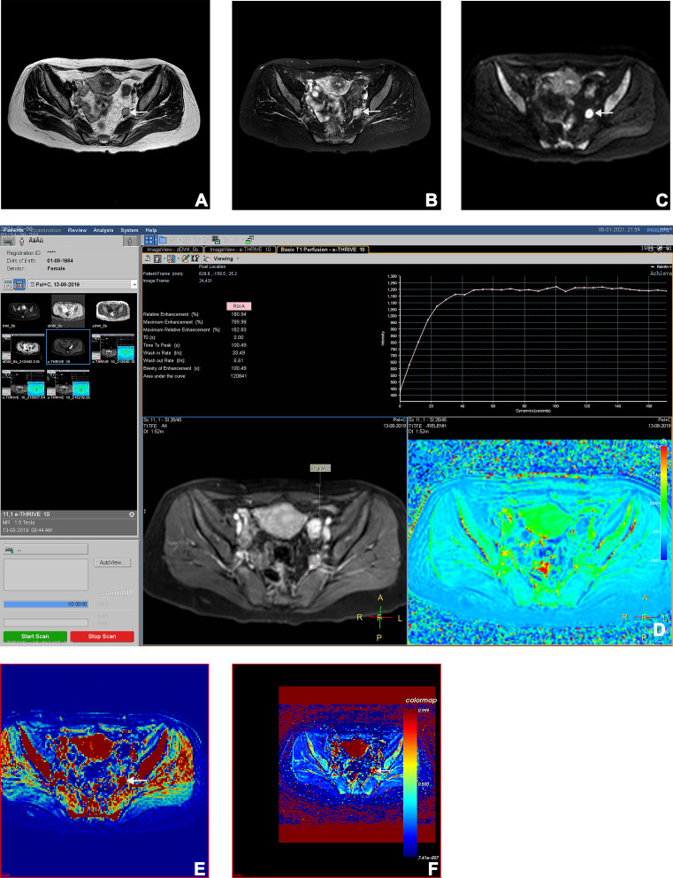
This retrospective study analyzed MRIs of 209 PLNs in 25 women with pathologically proven cervical cancer. All PLNs had been assessed by pre-treatment multimodal MRIs, and their status was finally confirmed by histopathology. In conventional MRI, lymph node characteristics were compared between metastatic and non-metastatic PLNs. Signal intensity, time-intensity curve (TIC) patterns minimal and mean apparent diffusion coefficients (ADC) were compared between them in DWI. In DCE-MRI, quantitative (K, K and V) analyses were performed on DCE-MRI sequences, and their predictive values were analyzed by ROC curves.

RESULTS

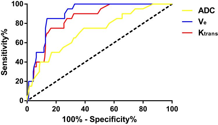
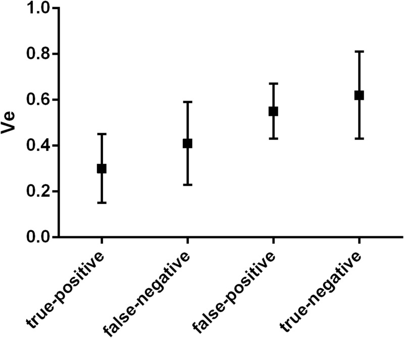
Of 209 PLNs, 22 (10.53%) were metastases and 187 (89.47%) were non-metastases at histopathologic examination. Considering a comparison of lymph node characteristics, the short axis size, the long axis size, and the boundary differed significantly between the two groups (<0.05).The differences in ADC, TIC types, K and V between metastatic and non-metastatic PLNs were significant as well (<0.05). The good diagnostic performance of multimodal MRI was shown in discriminating between metastatic and non-metastatic PLNs, with the sensitivity of 85.0% (17/20), specificity of 97.3% (184/189), and accuracy of 96.2% (201/209). ROC analyses showed that the diagnostic accuracy of ADC, K and V for discriminating between metastatic and non-metastatic PLNs in cervical cancer was 83.7%, 91.4%, and 92.4% with the cut-off values of 0.72 × 10mm/s, 0.52 min, and 0.53 min, respectively.

CONCLUSION

Multimodal MRI showed good diagnostic performance in determining PLN status in cervical cancer.

摘要

背景

盆腔淋巴结(PLN)转移状态影响宫颈癌患者的治疗和预后方案。然而,在临床实践中很难对其进行诊断。

目的

本研究旨在评估多模态磁共振成像(MRI)在鉴别宫颈癌转移性和非转移性盆腔淋巴结(PLN)中的诊断价值。

方法

这项回顾性研究分析了25例经病理证实为宫颈癌的女性患者的209个PLN的MRI图像。所有PLN均在治疗前通过多模态MRI进行评估,其状态最终通过组织病理学确认。在传统MRI中,比较转移性和非转移性PLN的淋巴结特征。在弥散加权成像(DWI)中,比较它们之间的信号强度、时间-强度曲线(TIC)类型、最小表观扩散系数(ADC)和平均表观扩散系数。在动态对比增强磁共振成像(DCE-MRI)中,对DCE-MRI序列进行定量(Ktrans、Kep和V)分析,并通过ROC曲线分析其预测价值。

结果

在209个PLN中,组织病理学检查显示22个(10.53%)为转移灶,187个(89.47%)为非转移灶。考虑淋巴结特征的比较,两组之间的短轴大小、长轴大小和边界存在显著差异(P<0.05)。转移性和非转移性PLN之间的ADC、TIC类型、Ktrans和Kep差异也具有统计学意义(P<0.05)。多模态MRI在鉴别转移性和非转移性PLN方面表现出良好的诊断性能,敏感性为85.0%(17/20),特异性为97.3%(184/189),准确性为96.2%(201/209)。ROC分析显示,ADC、Ktrans和Kep鉴别宫颈癌转移性和非转移性PLN的诊断准确性分别为83.7%、91.4%和92.4%,截断值分别为0.72×10⁻³mm²/s、0.52 min⁻¹和0.53 min⁻¹。

结论

多模态MRI在确定宫颈癌PLN状态方面表现出良好的诊断性能。

https://cdn.ncbi.nlm.nih.gov/pmc/blobs/2099/9326496/eff95e3d87e7/IJGM-15-6279-g0006.jpg
https://cdn.ncbi.nlm.nih.gov/pmc/blobs/2099/9326496/e594c5418f18/IJGM-15-6279-g0001.jpg
https://cdn.ncbi.nlm.nih.gov/pmc/blobs/2099/9326496/b6b907f684f3/IJGM-15-6279-g0002.jpg
https://cdn.ncbi.nlm.nih.gov/pmc/blobs/2099/9326496/0acd586b98c2/IJGM-15-6279-g0003.jpg
https://cdn.ncbi.nlm.nih.gov/pmc/blobs/2099/9326496/80918105b2ec/IJGM-15-6279-g0004.jpg
https://cdn.ncbi.nlm.nih.gov/pmc/blobs/2099/9326496/f319ec04f711/IJGM-15-6279-g0005.jpg
https://cdn.ncbi.nlm.nih.gov/pmc/blobs/2099/9326496/eff95e3d87e7/IJGM-15-6279-g0006.jpg

相似文献

1
Diagnostic Value of Multimodal Magnetic Resonance Imaging in Discriminating Between Metastatic and Non-Metastatic Pelvic Lymph Nodes in Cervical Cancer.多模态磁共振成像在鉴别宫颈癌盆腔转移性和非转移性淋巴结中的诊断价值
Int J Gen Med. 2022 Jul 22;15:6279-6288. doi: 10.2147/IJGM.S372154. eCollection 2022.
2
Diagnostic value of diffusion-weighted magnetic resonance imaging in discriminating between metastatic and non-metastatic pelvic lymph nodes in endometrial cancer.扩散加权磁共振成像在鉴别子宫内膜癌盆腔转移性与非转移性淋巴结中的诊断价值
Acta Radiol. 2020 Nov;61(11):1580-1586. doi: 10.1177/0284185120906660. Epub 2020 Feb 27.
3
Value of MRI and diffusion-weighted imaging in diagnosing normal-sized pelvic lymph nodes metastases in patients with cervical cancer.MRI 和弥散加权成像在诊断宫颈癌患者正常大小盆腔淋巴结转移中的价值。
Br J Radiol. 2022 Sep 1;95(1138):20200203. doi: 10.1259/bjr.20200203. Epub 2022 Aug 4.
4
Application of apparent diffusion coefficient values derived from diffusion-weighted imaging for assessing different sized metastatic lymph nodes in cervical cancers.基于扩散加权成像得出的表观扩散系数值在评估宫颈癌不同大小转移淋巴结中的应用。
Acta Radiol. 2020 Jun;61(6):848-855. doi: 10.1177/0284185119879686. Epub 2019 Oct 15.
5
The Role of Multiparametric Magnetic Resonance Imaging in the Study of Primary Tumor and Pelvic Lymph Node Metastasis in Stage IB1-IIA1 Cervical Cancer.多参数磁共振成像在IB1-IIA1期宫颈癌原发肿瘤及盆腔淋巴结转移研究中的作用
J Comput Assist Tomogr. 2020 Sep/Oct;44(5):750-758. doi: 10.1097/RCT.0000000000001084.
6
Diffusion-weighted magnetic resonance imaging of primary cervical cancer in the detection of sub-centimetre metastatic lymph nodes.原发性宫颈癌的弥散加权磁共振成像在检测亚厘米转移性淋巴结中的应用。
Cancer Imaging. 2020 Apr 6;20(1):27. doi: 10.1186/s40644-020-00303-4.
7
The value of multimodal magnetic resonance imaging in breast cancer and its correlation with pathological features and prognosis.多模态磁共振成像在乳腺癌中的价值及其与病理特征和预后的相关性。
Eur Rev Med Pharmacol Sci. 2023 Sep;27(18):8397-8403. doi: 10.26355/eurrev_202309_33762.
8
Assessment of pelvic lymph node metastasis in FIGO IB and IIA cervical cancer using quantitative dynamic contrast-enhanced MRI parameters.采用定量动态对比增强 MRI 参数评估 FIGO IB 和 IIA 期宫颈癌的盆腔淋巴结转移。
Diagn Interv Radiol. 2020 Sep;26(5):382-389. doi: 10.5152/dir.2020.19365.
9
A preliminary exploration of the intravoxel incoherent motion applied in the preoperative evaluation of mediastinal lymph node metastasis of lung cancer.体素内不相干运动在肺癌纵隔淋巴结转移术前评估中的初步探索。
J Thorac Dis. 2017 Apr;9(4):1073-1080. doi: 10.21037/jtd.2017.03.110.
10
Role of diffusion-weighted MRI: predicting axillary lymph node metastases in breast cancer.扩散加权磁共振成像的作用:预测乳腺癌腋窝淋巴结转移
Acta Radiol. 2014 Oct;55(8):909-16. doi: 10.1177/0284185113509094. Epub 2013 Nov 14.

引用本文的文献

1
Tumor markers and multimodal magnetic resonance imaging in predicting rectal cancer stage and differentiation.肿瘤标志物与多模态磁共振成像在预测直肠癌分期及分化程度中的应用
World J Gastrointest Oncol. 2025 Aug 15;17(8):108007. doi: 10.4251/wjgo.v17.i8.108007.
2
The value of multiparameter MRI of early cervical cancer combined with SCC-Ag in predicting its pelvic lymph node metastasis.早期宫颈癌多参数磁共振成像联合鳞状细胞癌抗原预测盆腔淋巴结转移的价值。
Front Oncol. 2024 Sep 11;14:1417933. doi: 10.3389/fonc.2024.1417933. eCollection 2024.
3
Machine Learning-Based Multiparametric Magnetic Resonance Imaging Radiomics Model for Preoperative Predicting the Deep Stromal Invasion in Patients with Early Cervical Cancer.

本文引用的文献

1
A comparison of the multimodal magnetic resonance imaging features of brain metastases vs. high-grade gliomas.脑转移瘤与高级别胶质瘤的多模态磁共振成像特征比较。
Am J Transl Res. 2021 Apr 15;13(4):3543-3548. eCollection 2021.
2
Application of apparent diffusion coefficient values derived from diffusion-weighted imaging for assessing different sized metastatic lymph nodes in cervical cancers.基于扩散加权成像得出的表观扩散系数值在评估宫颈癌不同大小转移淋巴结中的应用。
Acta Radiol. 2020 Jun;61(6):848-855. doi: 10.1177/0284185119879686. Epub 2019 Oct 15.
3
Diagnostic performance of magnetic resonance to assess treatment response after neoadjuvant therapy in patients with locally advanced rectal cancer.
基于机器学习的多参数磁共振成像放射组学模型在预测早期宫颈癌患者深层间质浸润中的应用。
J Imaging Inform Med. 2024 Feb;37(1):230-246. doi: 10.1007/s10278-023-00906-w. Epub 2024 Jan 10.
4
Prognostic analysis of patients with stage IIIC1p cervical cancer treated by surgery.ⅡIC1p 期宫颈癌患者行手术治疗的预后分析。
World J Surg Oncol. 2023 Jun 21;21(1):186. doi: 10.1186/s12957-023-03076-9.
磁共振成像评估局部进展期直肠癌新辅助治疗后治疗反应的诊断性能。
Abdom Radiol (NY). 2019 Nov;44(11):3632-3640. doi: 10.1007/s00261-019-01894-8.
4
Cervical cancer.宫颈癌。
Lancet. 2019 Jan 12;393(10167):169-182. doi: 10.1016/S0140-6736(18)32470-X.
5
Comparison of the prognostic value of F-18 FDG PET/CT metabolic parameters of primary tumors and MRI findings in patients with locally advanced cervical cancer treated with concurrent chemoradiotherapy.同步放化疗治疗的局部晚期宫颈癌患者中原发性肿瘤的F-18 FDG PET/CT代谢参数与MRI表现的预后价值比较
Brachytherapy. 2019 Mar-Apr;18(2):154-162. doi: 10.1016/j.brachy.2018.11.005. Epub 2018 Dec 26.
6
Validation of the 2018 FIGO cervical cancer staging system.FIGO 2018 宫颈癌分期系统的验证。
Gynecol Oncol. 2019 Jan;152(1):87-93. doi: 10.1016/j.ygyno.2018.10.026. Epub 2018 Oct 30.
7
Role of Quantitative Dynamic Contrast-Enhanced MRI in Evaluating Regional Lymph Nodes With a Short-Axis Diameter of Less Than 5 mm in Rectal Cancer.定量动态对比增强 MRI 在评估直肠癌短轴直径小于 5mm 的区域淋巴结中的作用。
AJR Am J Roentgenol. 2019 Jan;212(1):77-83. doi: 10.2214/AJR.18.19866. Epub 2018 Oct 24.
8
Diffusion MRI of cancer: From low to high b-values.癌症的扩散 MRI:从低到高 b 值。
J Magn Reson Imaging. 2019 Jan;49(1):23-40. doi: 10.1002/jmri.26293. Epub 2018 Oct 12.
9
Contrast agents in dynamic contrast-enhanced magnetic resonance imaging.动态对比增强磁共振成像中的对比剂
Oncotarget. 2017 Jun 27;8(26):43491-43505. doi: 10.18632/oncotarget.16482.
10
A Comprehensive Comparison of CT, MRI, Positron Emission Tomography or Positron Emission Tomography/CT, and Diffusion Weighted Imaging-MRI for Detecting the Lymph Nodes Metastases in Patients with Cervical Cancer: A Meta-Analysis Based on 67 Studies.CT、MRI、正电子发射断层扫描或正电子发射断层扫描/CT以及扩散加权成像-MRI检测宫颈癌患者淋巴结转移的综合比较:基于67项研究的荟萃分析
Gynecol Obstet Invest. 2017;82(3):209-222. doi: 10.1159/000456006. Epub 2017 Feb 10.