• 文献检索
  • 文档翻译
  • 深度研究
  • 学术资讯
  • Suppr Zotero 插件Zotero 插件
  • 邀请有礼
  • 套餐&价格
  • 历史记录
应用&插件
Suppr Zotero 插件Zotero 插件浏览器插件Mac 客户端Windows 客户端微信小程序
定价
高级版会员购买积分包购买API积分包
服务
文献检索文档翻译深度研究API 文档MCP 服务
关于我们
关于 Suppr公司介绍联系我们用户协议隐私条款
关注我们

Suppr 超能文献

核心技术专利:CN118964589B侵权必究
粤ICP备2023148730 号-1Suppr @ 2026

文献检索

告别复杂PubMed语法,用中文像聊天一样搜索,搜遍4000万医学文献。AI智能推荐,让科研检索更轻松。

立即免费搜索

文件翻译

保留排版,准确专业,支持PDF/Word/PPT等文件格式,支持 12+语言互译。

免费翻译文档

深度研究

AI帮你快速写综述,25分钟生成高质量综述,智能提取关键信息,辅助科研写作。

立即免费体验

表皮生长因子受体配体通过上调 BIN3 抑制侵袭,将 EGFR 扩增型胶质母细胞瘤中 EGFR 的致癌基因角色转变为肿瘤抑制基因。

EGFR ligand shifts the role of EGFR from oncogene to tumour suppressor in EGFR-amplified glioblastoma by suppressing invasion through BIN3 upregulation.

机构信息

Department of Neurology, University of Texas Southwestern Medical Center, Dallas, TX, USA.

Hubei Province Key Laboratory of Allergy and Immunology and Department of Immunology, School of Basic Medical Sciences, Taikang Medical School, Wuhan University, Wuhan, China.

出版信息

Nat Cell Biol. 2022 Aug;24(8):1291-1305. doi: 10.1038/s41556-022-00962-4. Epub 2022 Aug 1.

DOI:10.1038/s41556-022-00962-4
PMID:
35915159
原文链接:https://pmc.ncbi.nlm.nih.gov/articles/PMC9389625/
Abstract

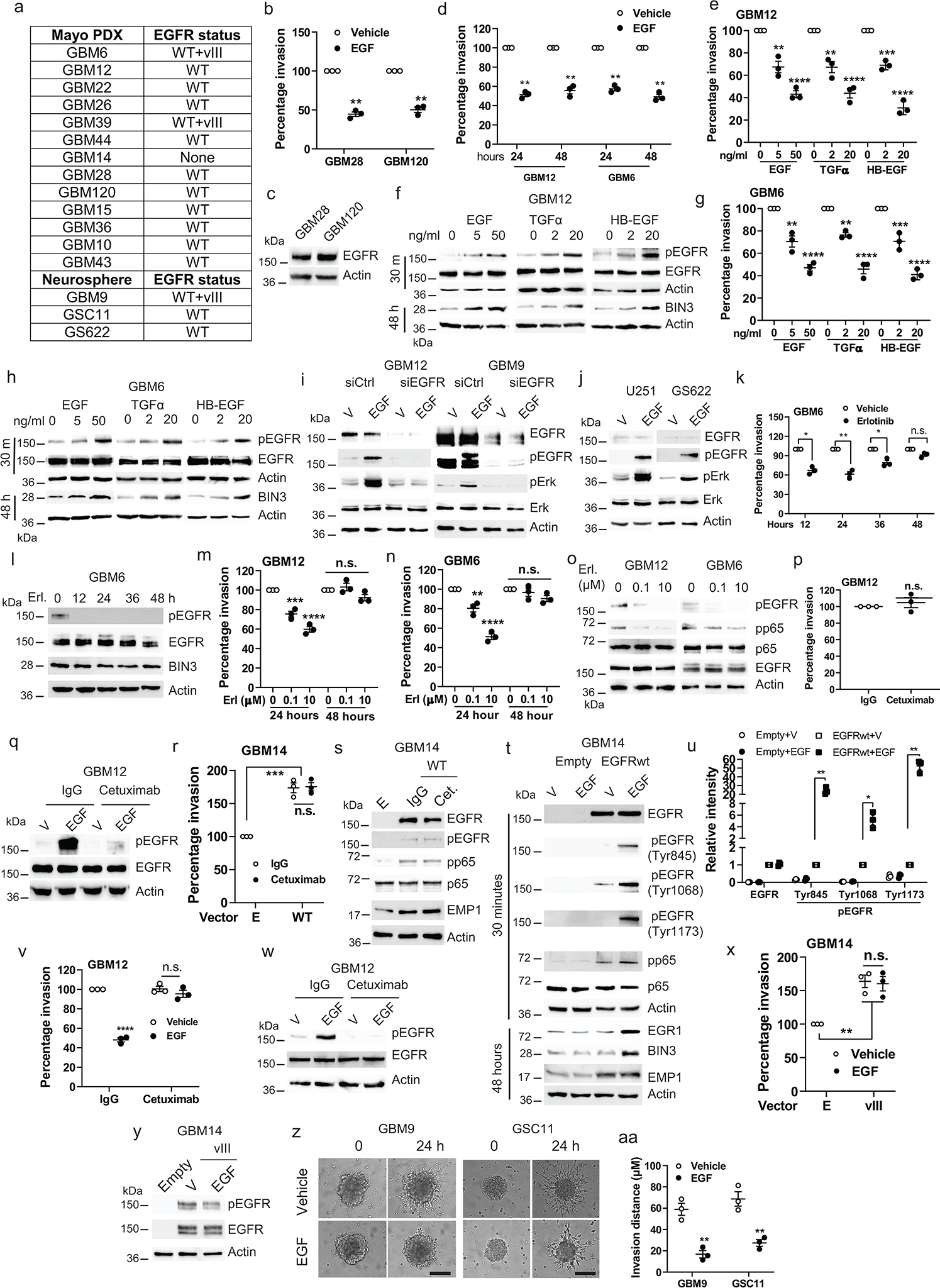
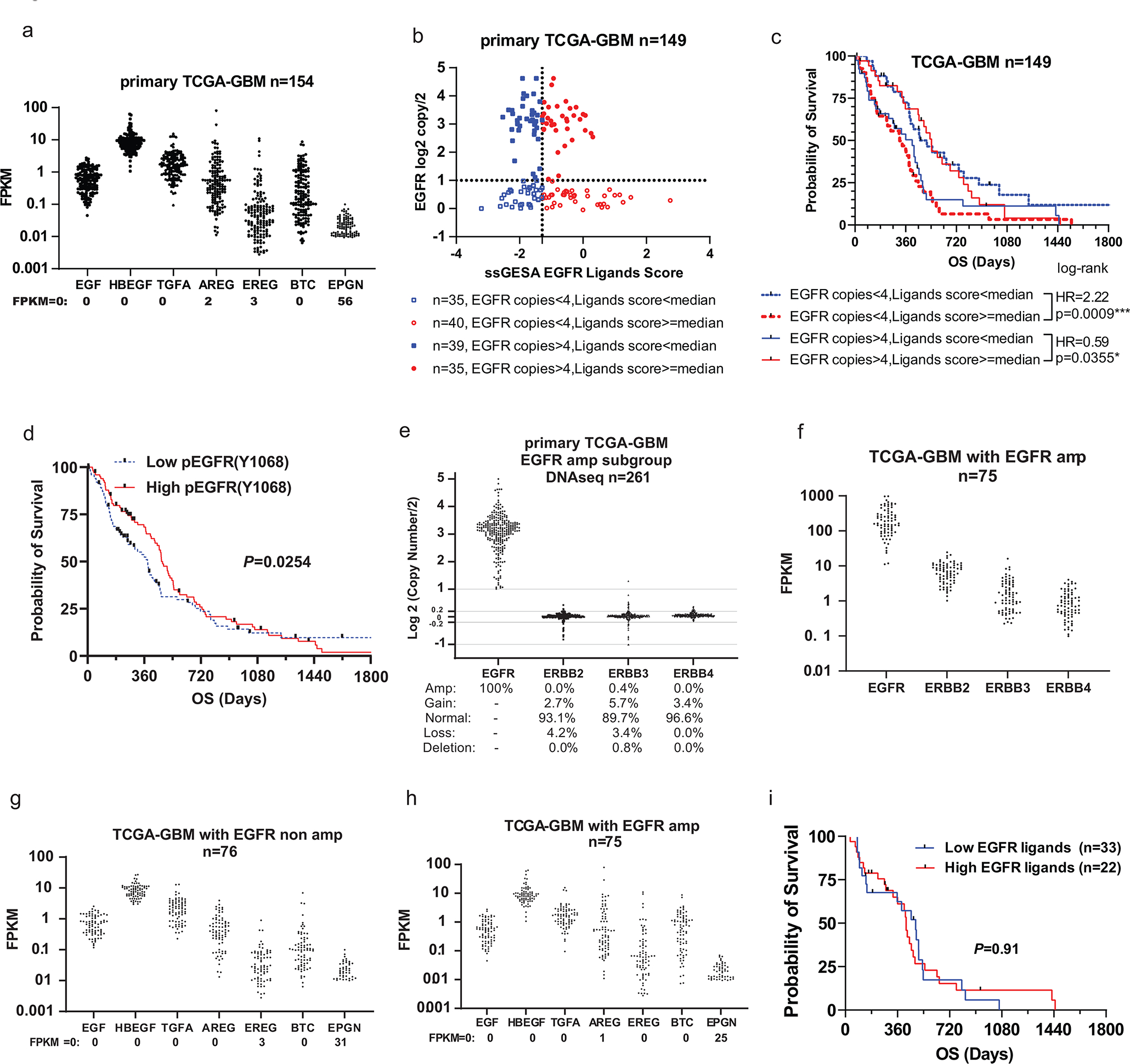
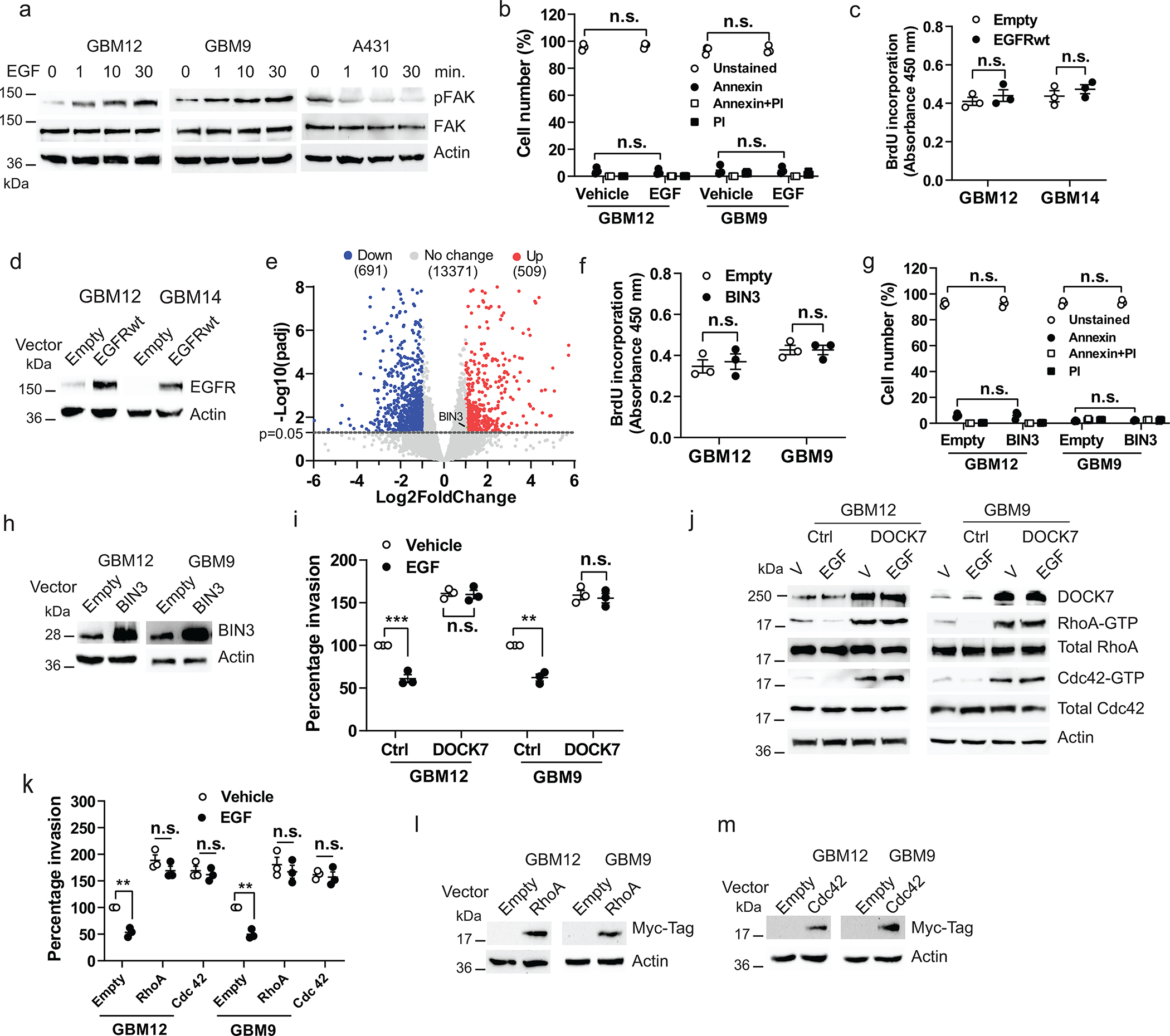
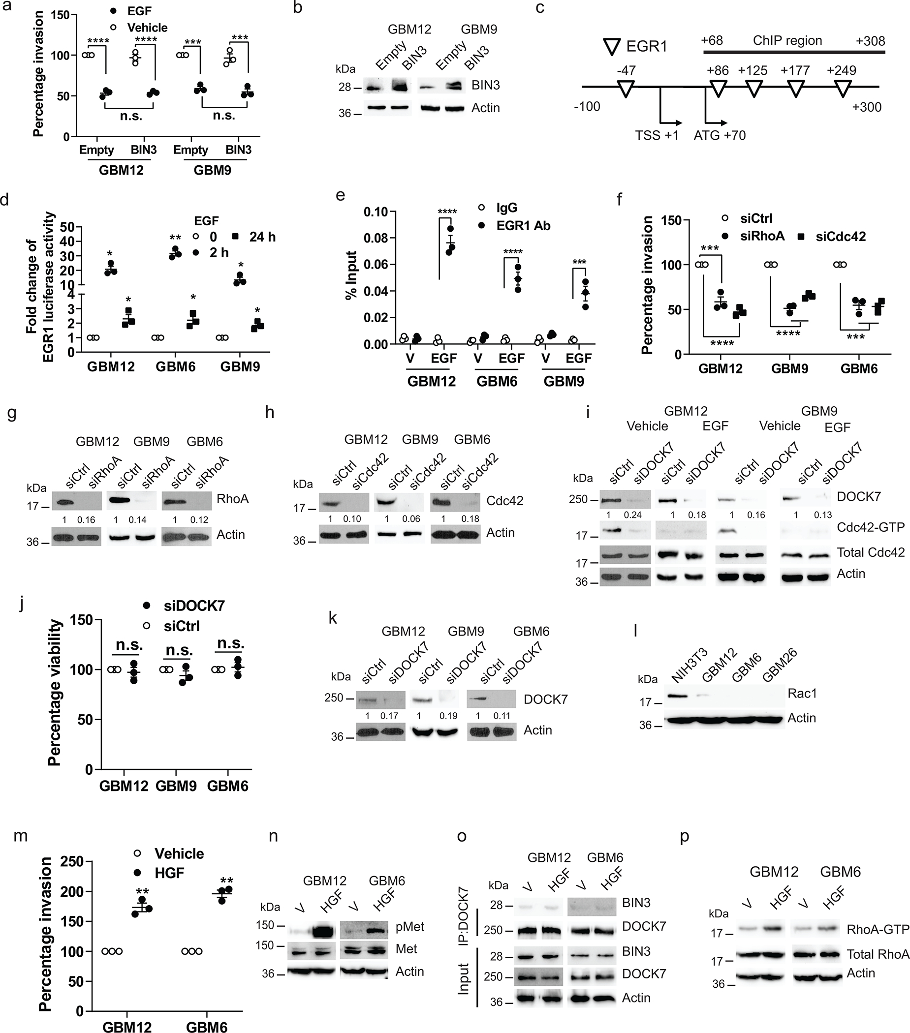
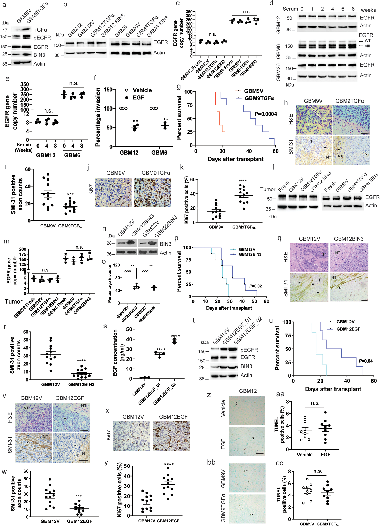
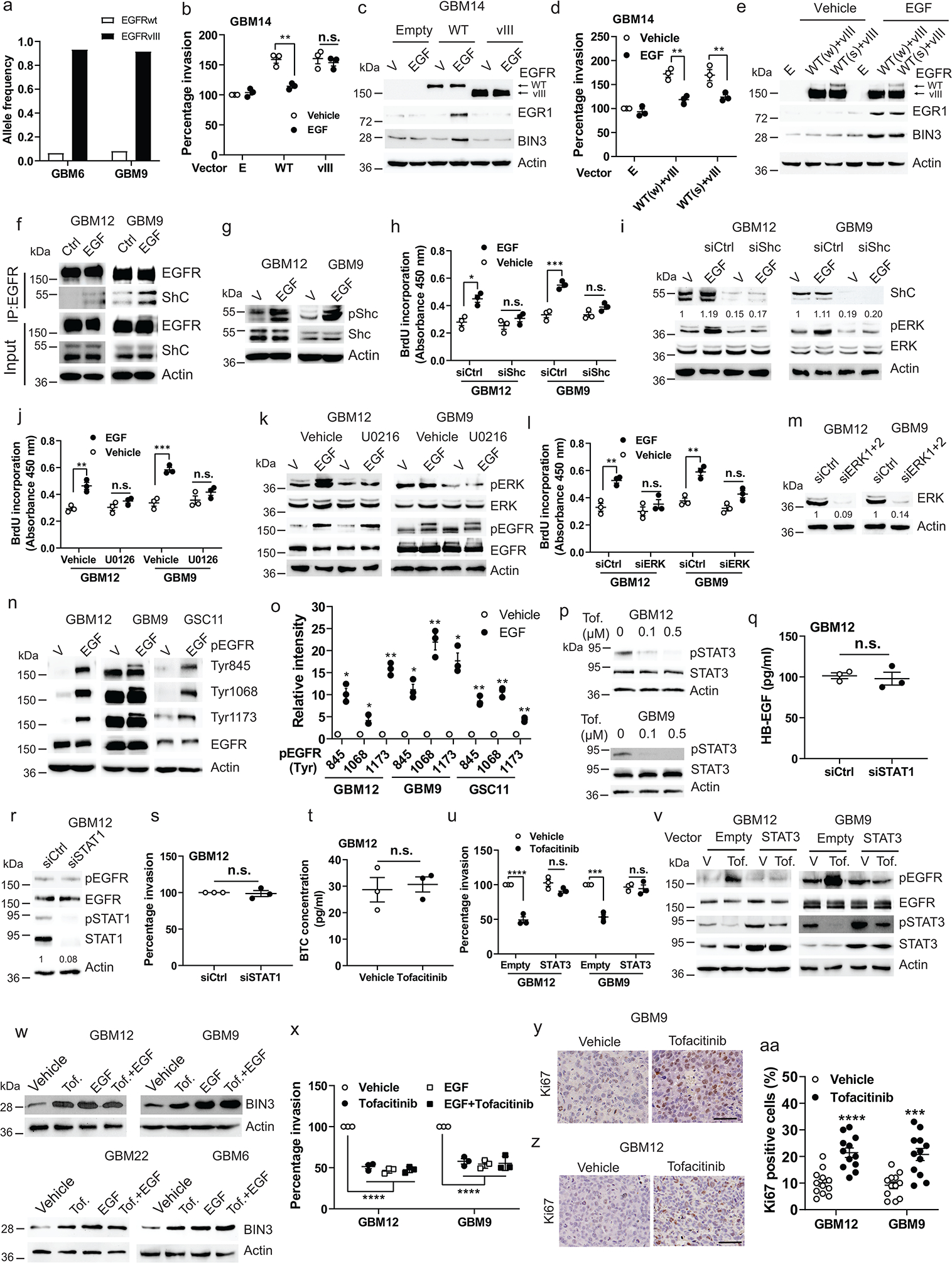
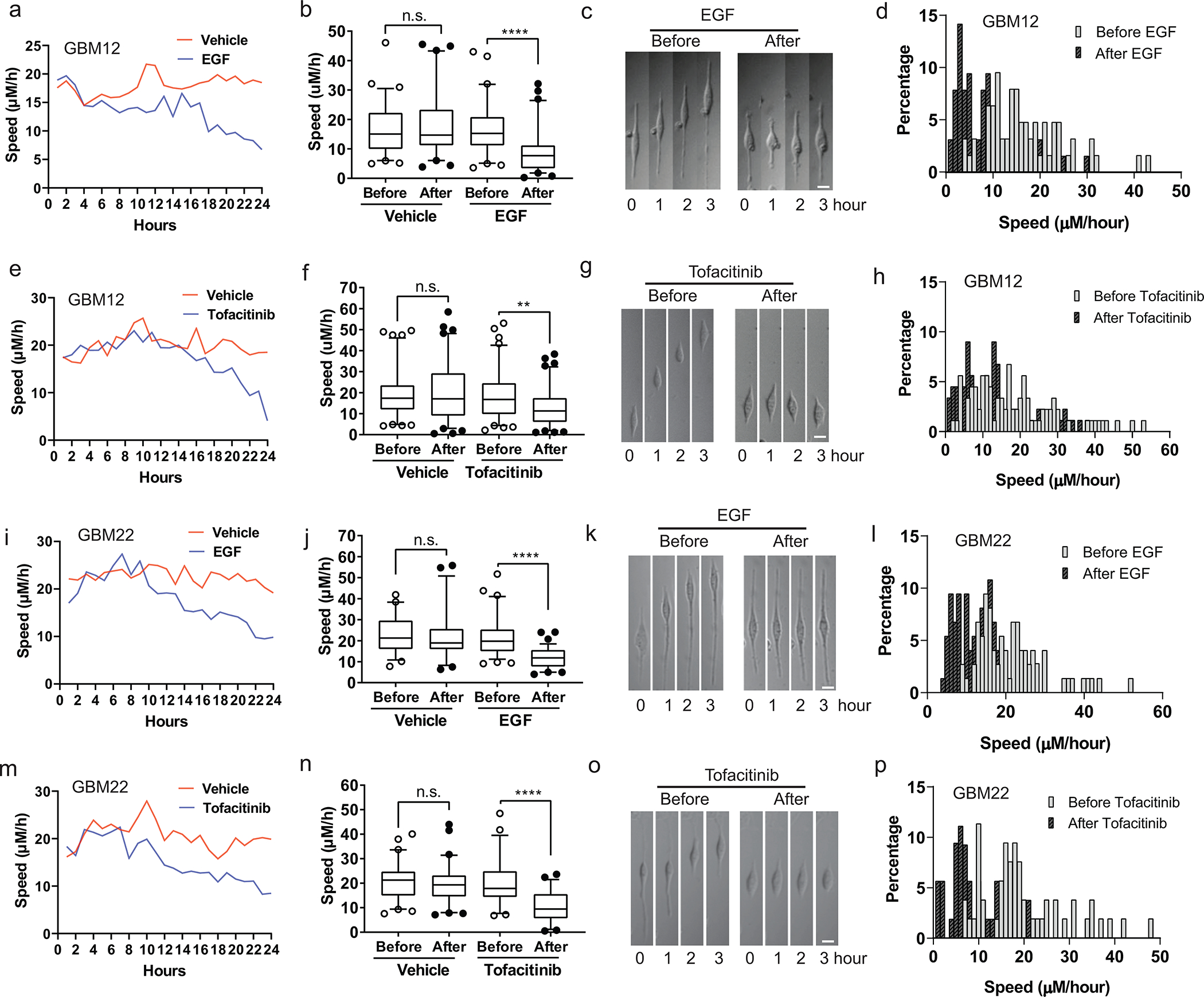
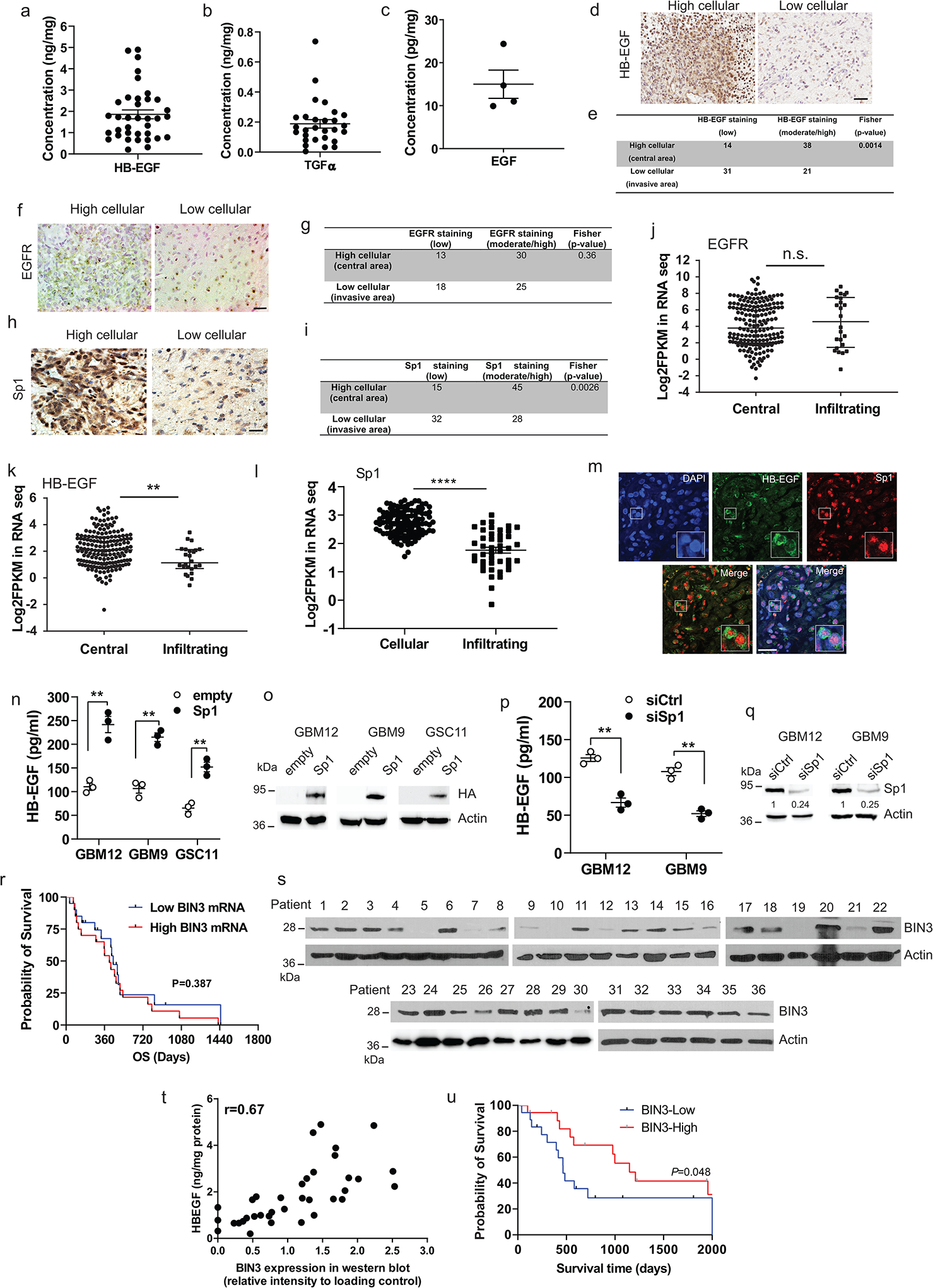
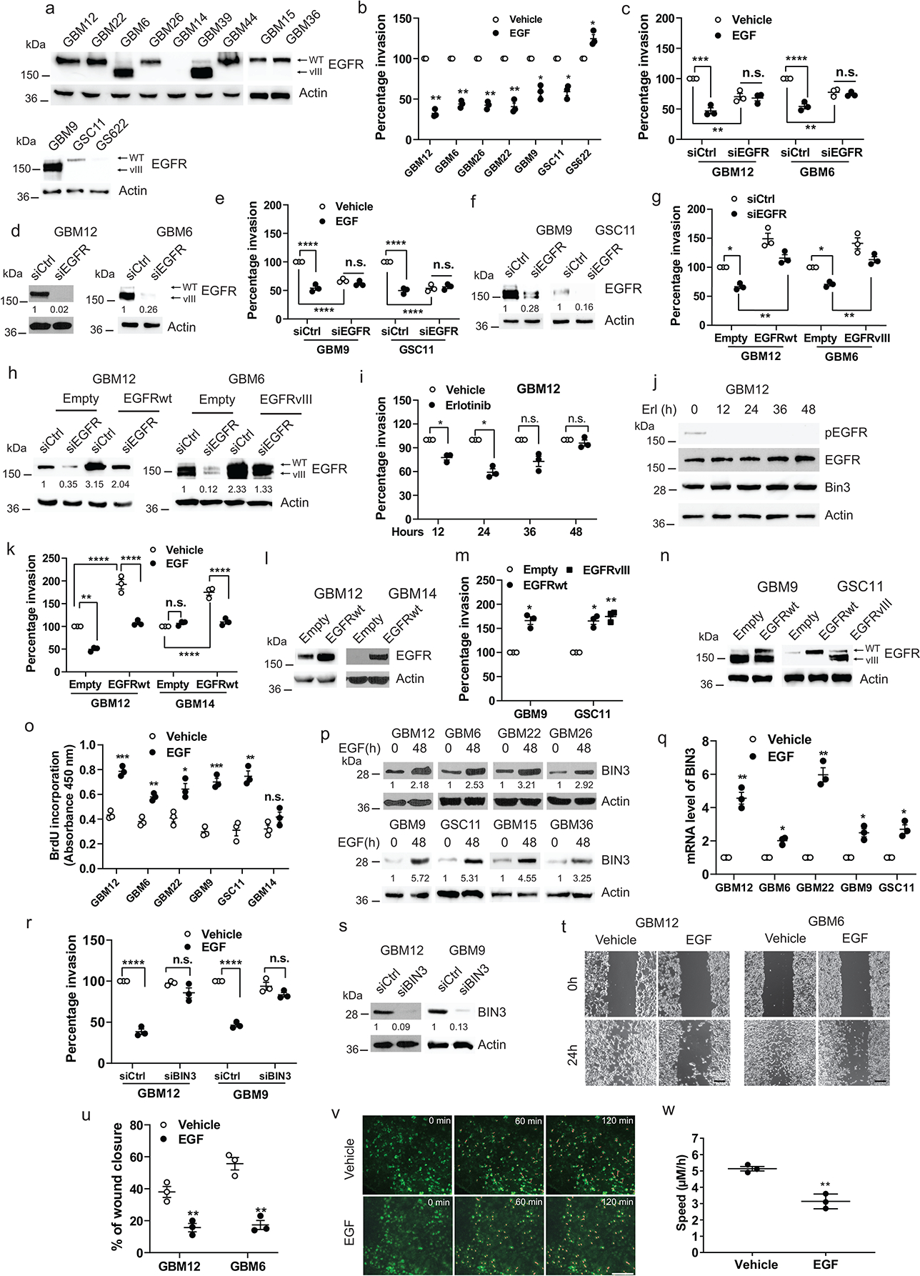
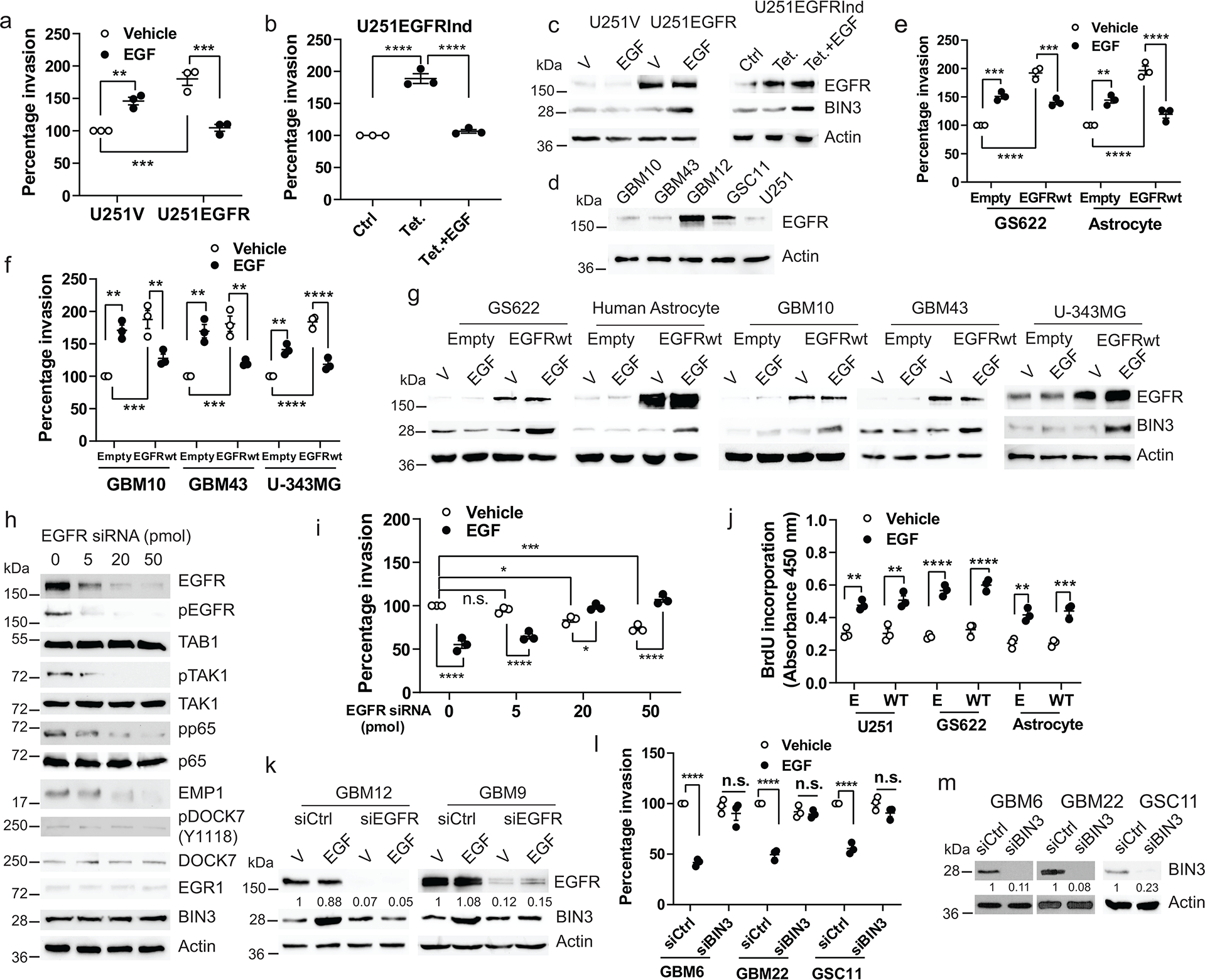
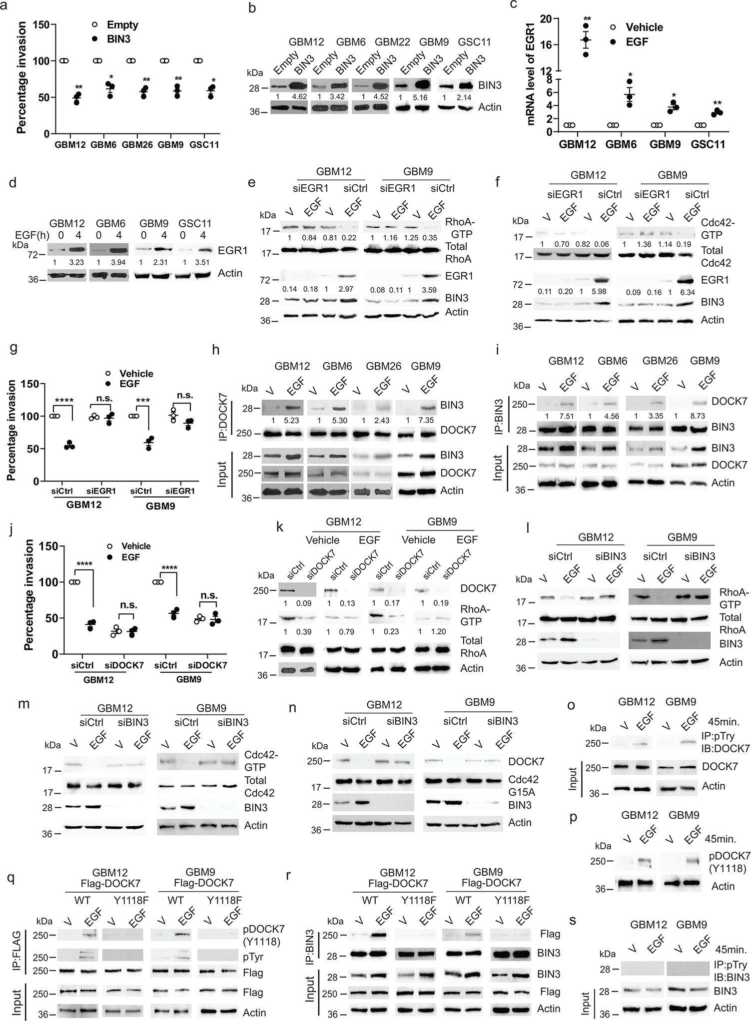
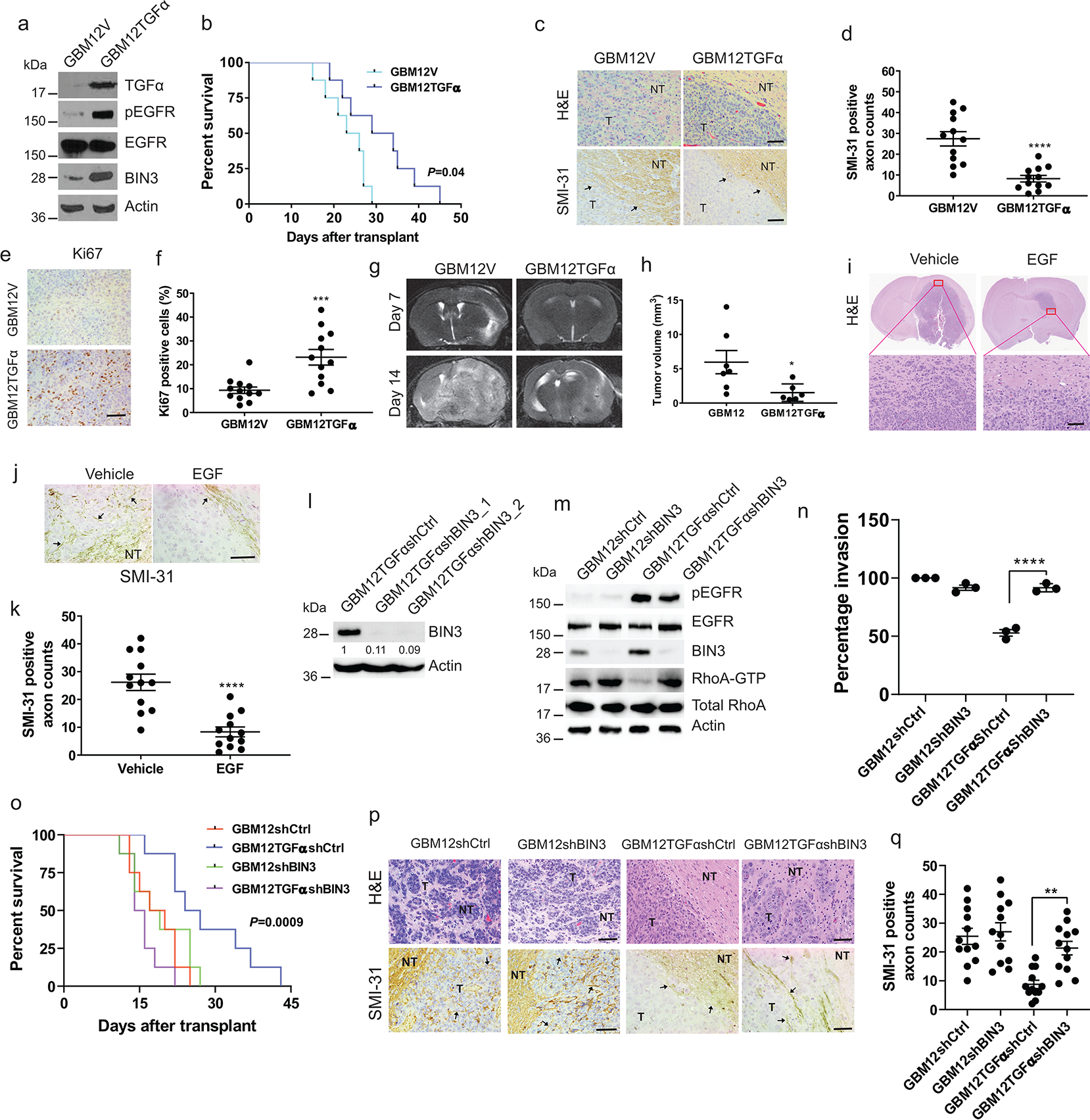
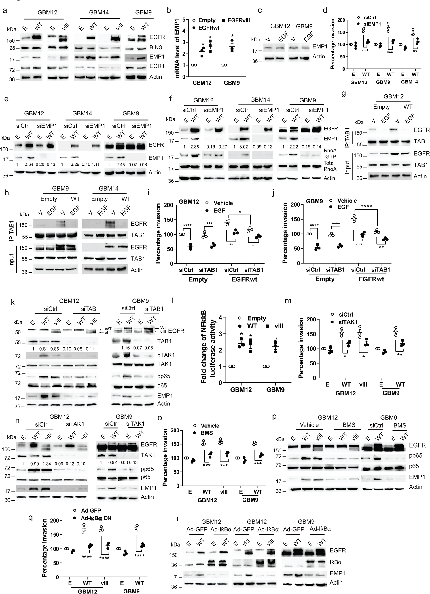
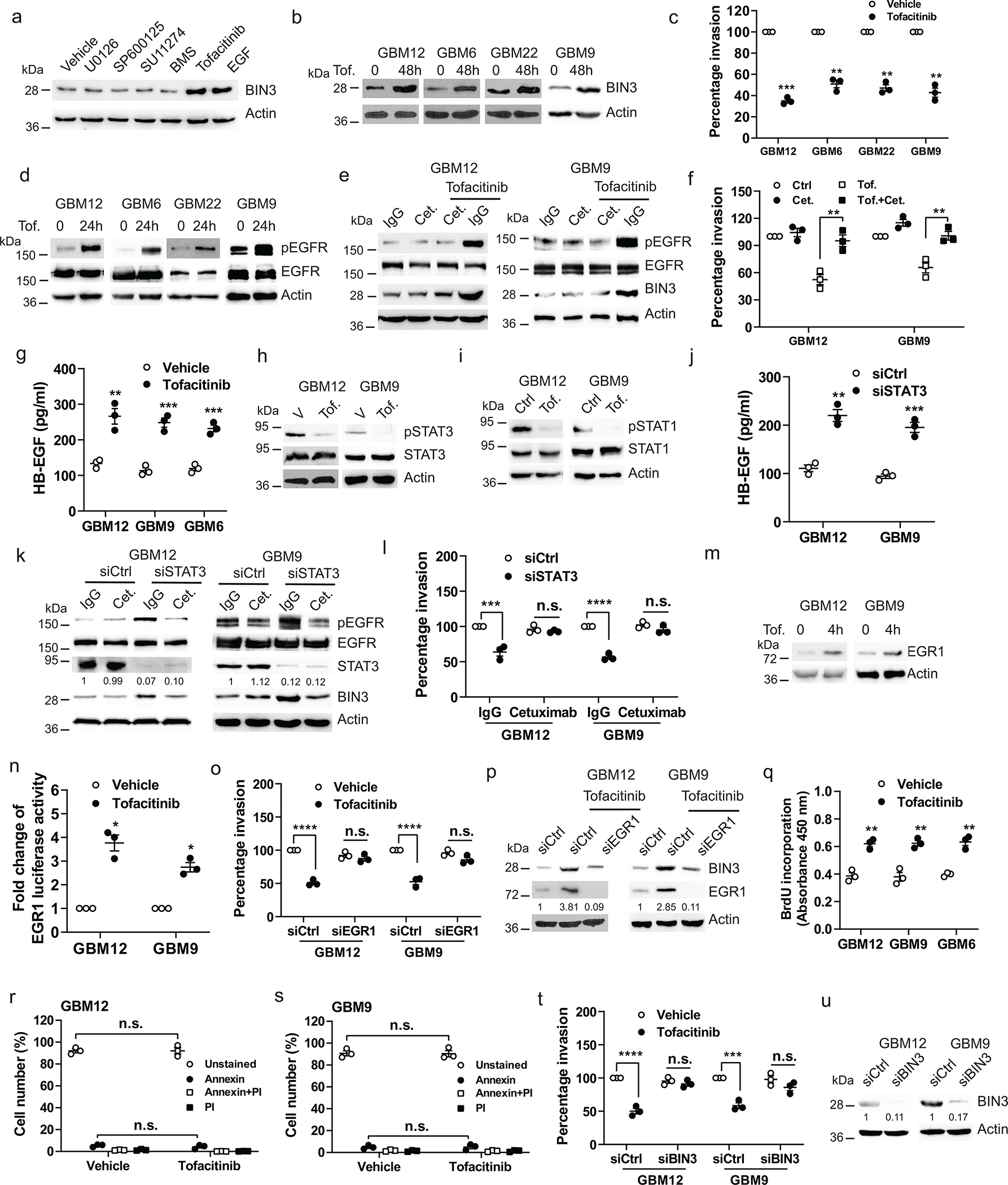
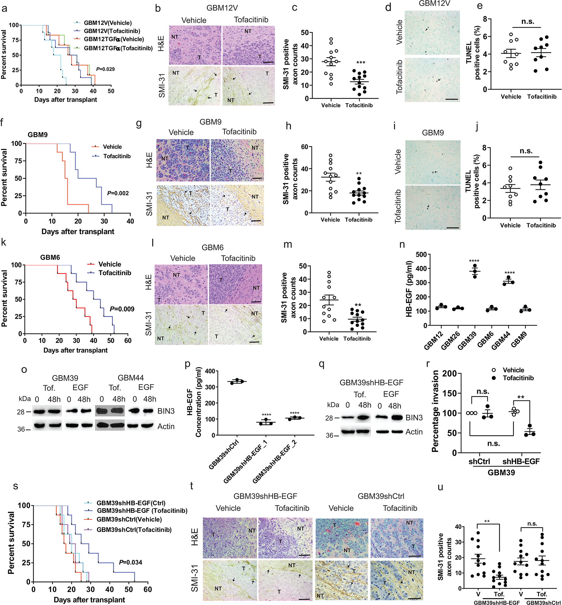
The epidermal growth factor receptor (EGFR) is a prime oncogene that is frequently amplified in glioblastomas. Here we demonstrate a new tumour-suppressive function of EGFR in EGFR-amplified glioblastomas regulated by EGFR ligands. Constitutive EGFR signalling promotes invasion via activation of a TAB1-TAK1-NF-κB-EMP1 pathway, resulting in large tumours and decreased survival in orthotopic models. Ligand-activated EGFR promotes proliferation and surprisingly suppresses invasion by upregulating BIN3, which inhibits a DOCK7-regulated Rho GTPase pathway, resulting in small hyperproliferating non-invasive tumours and improved survival. Data from The Cancer Genome Atlas reveal that in EGFR-amplified glioblastomas, a low level of EGFR ligands confers a worse prognosis, whereas a high level of EGFR ligands confers an improved prognosis. Thus, increased EGFR ligand levels shift the role of EGFR from oncogene to tumour suppressor in EGFR-amplified glioblastomas by suppressing invasion. The tumour-suppressive function of EGFR can be activated therapeutically using tofacitinib, which suppresses invasion by increasing EGFR ligand levels and upregulating BIN3.

摘要

表皮生长因子受体(EGFR)是一种主要的致癌基因,在胶质母细胞瘤中经常扩增。在这里,我们证明了 EGFR 配体调控的 EGFR 扩增胶质母细胞瘤中 EGFR 的一个新的肿瘤抑制功能。组成性 EGFR 信号通过激活 TAB1-TAK1-NF-κB-EMP1 途径促进侵袭,导致肿瘤体积增大和原位模型中生存率降低。配体激活的 EGFR 通过上调 BIN3 促进增殖,令人惊讶的是抑制了 DOCK7 调节的 Rho GTPase 途径,导致小的过度增殖性非侵袭性肿瘤和生存率提高。来自癌症基因组图谱的数据表明,在 EGFR 扩增的胶质母细胞瘤中,低水平的 EGFR 配体预示着更差的预后,而高水平的 EGFR 配体预示着更好的预后。因此,增加 EGFR 配体水平通过抑制侵袭将 EGFR 的作用从致癌基因转变为 EGFR 扩增胶质母细胞瘤中的肿瘤抑制因子。使用托法替尼可以激活 EGFR 的肿瘤抑制功能,托法替尼通过增加 EGFR 配体水平和上调 BIN3 来抑制侵袭。

https://cdn.ncbi.nlm.nih.gov/pmc/blobs/21ba/9389625/ac05491070bd/nihms-1824714-f0008.jpg
https://cdn.ncbi.nlm.nih.gov/pmc/blobs/21ba/9389625/2cc862e9f775/nihms-1824714-f0009.jpg
https://cdn.ncbi.nlm.nih.gov/pmc/blobs/21ba/9389625/af4f71173823/nihms-1824714-f0010.jpg
https://cdn.ncbi.nlm.nih.gov/pmc/blobs/21ba/9389625/898ce96fddca/nihms-1824714-f0011.jpg
https://cdn.ncbi.nlm.nih.gov/pmc/blobs/21ba/9389625/bbfef098f229/nihms-1824714-f0012.jpg
https://cdn.ncbi.nlm.nih.gov/pmc/blobs/21ba/9389625/7dbc8ef09c70/nihms-1824714-f0013.jpg
https://cdn.ncbi.nlm.nih.gov/pmc/blobs/21ba/9389625/9261ecdc51ee/nihms-1824714-f0014.jpg
https://cdn.ncbi.nlm.nih.gov/pmc/blobs/21ba/9389625/144424678452/nihms-1824714-f0015.jpg
https://cdn.ncbi.nlm.nih.gov/pmc/blobs/21ba/9389625/efedc2e92c7b/nihms-1824714-f0001.jpg
https://cdn.ncbi.nlm.nih.gov/pmc/blobs/21ba/9389625/098ba8ea2e0d/nihms-1824714-f0002.jpg
https://cdn.ncbi.nlm.nih.gov/pmc/blobs/21ba/9389625/a42f03140d7d/nihms-1824714-f0003.jpg
https://cdn.ncbi.nlm.nih.gov/pmc/blobs/21ba/9389625/aeabc6f764c7/nihms-1824714-f0004.jpg
https://cdn.ncbi.nlm.nih.gov/pmc/blobs/21ba/9389625/374bc8841fa9/nihms-1824714-f0005.jpg
https://cdn.ncbi.nlm.nih.gov/pmc/blobs/21ba/9389625/802b6be02796/nihms-1824714-f0006.jpg
https://cdn.ncbi.nlm.nih.gov/pmc/blobs/21ba/9389625/1e02098151ec/nihms-1824714-f0007.jpg
https://cdn.ncbi.nlm.nih.gov/pmc/blobs/21ba/9389625/ac05491070bd/nihms-1824714-f0008.jpg

相似文献

1
EGFR ligand shifts the role of EGFR from oncogene to tumour suppressor in EGFR-amplified glioblastoma by suppressing invasion through BIN3 upregulation.表皮生长因子受体配体通过上调 BIN3 抑制侵袭,将 EGFR 扩增型胶质母细胞瘤中 EGFR 的致癌基因角色转变为肿瘤抑制基因。
Nat Cell Biol. 2022 Aug;24(8):1291-1305. doi: 10.1038/s41556-022-00962-4. Epub 2022 Aug 1.
2
Monosomy of chromosome 10 associated with dysregulation of epidermal growth factor signaling in glioblastomas.10号染色体单体与胶质母细胞瘤中表皮生长因子信号传导失调相关。
JAMA. 2009 Jul 15;302(3):276-89. doi: 10.1001/jama.2009.1022.
3
MicroRNA-566 activates EGFR signaling and its inhibition sensitizes glioblastoma cells to nimotuzumab.微小RNA-566激活表皮生长因子受体信号通路,抑制该因子可使胶质母细胞瘤细胞对尼妥珠单抗敏感。
Mol Cancer. 2014 Mar 20;13:63. doi: 10.1186/1476-4598-13-63.
4
Epidermal growth factor receptor and ligand family expression and activity in glioblastoma.表皮生长因子受体及其配体家族在胶质母细胞瘤中的表达和活性。
J Neurochem. 2018 Oct;147(1):99-109. doi: 10.1111/jnc.14538. Epub 2018 Aug 16.
5
A Novel Signaling Complex between TROY and EGFR Mediates Glioblastoma Cell Invasion.TROY 和 EGFR 之间的新型信号复合物介导电胶质瘤细胞侵袭。
Mol Cancer Res. 2018 Feb;16(2):322-332. doi: 10.1158/1541-7786.MCR-17-0454. Epub 2017 Nov 8.
6
Inhibition of epidermal growth factor receptor suppresses parathyroid hormone-related protein expression in tumours and ameliorates cancer-associated cachexia.抑制表皮生长因子受体可抑制肿瘤中甲状旁腺激素相关蛋白的表达,并改善癌症相关性恶病质。
J Cachexia Sarcopenia Muscle. 2022 Jun;13(3):1582-1594. doi: 10.1002/jcsm.12985. Epub 2022 Apr 4.
7
Distinct domains in the SHP-2 phosphatase differentially regulate epidermal growth factor receptor/NF-kappaB activation through Gab1 in glioblastoma cells.SHP-2磷酸酶中的不同结构域通过胶质母细胞瘤细胞中的Gab1差异调节表皮生长因子受体/NF-κB激活。
Mol Cell Biol. 2004 Jan;24(2):823-36. doi: 10.1128/MCB.24.2.823-836.2004.
8
Androgen Receptor Activation in Glioblastoma Can Be Achieved by Ligand-Independent Signaling through EGFR-A Potential Therapeutic Target.通过 EGFR 配体非依赖性信号转导实现胶质母细胞瘤中的雄激素受体激活——一种潜在的治疗靶点。
Int J Mol Sci. 2021 Oct 11;22(20):10954. doi: 10.3390/ijms222010954.
9
Oncogenic EGFR Represses the TET1 DNA Demethylase to Induce Silencing of Tumor Suppressors in Cancer Cells.致癌性表皮生长因子受体抑制TET1 DNA去甲基化酶以诱导癌细胞中肿瘤抑制基因沉默。
Cell Rep. 2016 Jul 12;16(2):457-471. doi: 10.1016/j.celrep.2016.05.087. Epub 2016 Jun 23.
10
Epidermal growth factor (EGF)-enhanced vascular cell adhesion molecule-1 (VCAM-1) expression promotes macrophage and glioblastoma cell interaction and tumor cell invasion.表皮生长因子(EGF)增强的血管细胞黏附分子-1(VCAM-1)表达促进巨噬细胞和神经胶质瘤细胞的相互作用和肿瘤细胞的侵袭。
J Biol Chem. 2013 Nov 1;288(44):31488-95. doi: 10.1074/jbc.M113.499020. Epub 2013 Sep 17.

引用本文的文献

1
Proteomics on choroidal neovascularization based on itraq and the protective effect of TAB1 in CNV.基于iTRAQ技术的脉络膜新生血管蛋白质组学及TAB1在脉络膜新生血管中的保护作用
Sci Rep. 2025 Aug 23;15(1):31037. doi: 10.1038/s41598-025-15134-1.
2
Precision Neuro-Oncology in Glioblastoma: AI-Guided CRISPR Editing and Real-Time Multi-Omics for Genomic Brain Surgery.胶质母细胞瘤中的精准神经肿瘤学:用于基因组脑手术的人工智能引导的CRISPR编辑和实时多组学技术
Int J Mol Sci. 2025 Jul 30;26(15):7364. doi: 10.3390/ijms26157364.
3
Decoding EGFR A289 Mutation in Glioblastoma: A Predictive Biomarker Framework and Targeted Therapeutic Insights.

本文引用的文献

1
Glioblastoma mutations alter EGFR dimer structure to prevent ligand bias.胶质母细胞瘤突变改变 EGFR 二聚体结构以防止配体偏向性。
Nature. 2022 Feb;602(7897):518-522. doi: 10.1038/s41586-021-04393-3. Epub 2022 Feb 9.
2
Comprehensive targeting of resistance to inhibition of RTK signaling pathways by using glucocorticoids.利用糖皮质激素全面靶向抑制 RTK 信号通路的耐药性。
Nat Commun. 2021 Dec 1;12(1):7014. doi: 10.1038/s41467-021-27276-7.
3
EGFR inhibition triggers an adaptive response by co-opting antiviral signaling pathways in lung cancer.
解码胶质母细胞瘤中的表皮生长因子受体(EGFR)A289突变:一个预测性生物标志物框架及靶向治疗见解
J Mol Neurosci. 2025 Jul 12;75(3):88. doi: 10.1007/s12031-025-02381-0.
4
Antibody-Drug Conjugates (ADCs): current and future biopharmaceuticals.抗体药物偶联物(ADCs):当前及未来的生物制药
J Hematol Oncol. 2025 Apr 30;18(1):51. doi: 10.1186/s13045-025-01704-3.
5
Transcriptomic analysis of co-expression and activation in glioblastoma reveals associations with its ligands.胶质母细胞瘤中共表达与激活的转录组分析揭示了其与配体的关联。
Neurooncol Adv. 2024 Dec 28;7(1):vdae229. doi: 10.1093/noajnl/vdae229. eCollection 2025 Jan-Dec.
6
Metabolomics and network pharmacology-based identification of phenolic acids in Polygonatum kingianum var. grandifolium rhizomes as anti-cancer/Tumor active ingredients.基于代谢组学和网络药理学鉴定滇黄精变种大叶黄精根茎中的酚酸作为抗癌/肿瘤活性成分
PLoS One. 2024 Dec 17;19(12):e0315857. doi: 10.1371/journal.pone.0315857. eCollection 2024.
7
The role of miR-152 in urological tumors: potential biomarkers and therapeutic targets.miR-152 在尿路上皮肿瘤中的作用:潜在的生物标志物和治疗靶点。
Front Immunol. 2024 Nov 13;15:1464327. doi: 10.3389/fimmu.2024.1464327. eCollection 2024.
8
Epithelial membrane protein 1 in human cancer: a potential diagnostic biomarker and therapeutic target.人癌症中的上皮膜蛋白1:一种潜在的诊断生物标志物和治疗靶点。
Biomark Med. 2024;18(21-22):995-1005. doi: 10.1080/17520363.2024.2416887. Epub 2024 Oct 29.
9
Exploring the prognostic value and biological pathways of transcriptomics and radiomics patterns in glioblastoma multiforme.探索多形性胶质母细胞瘤中转录组学和放射组学模式的预后价值及生物学途径。
Heliyon. 2024 Jun 27;10(13):e33760. doi: 10.1016/j.heliyon.2024.e33760. eCollection 2024 Jul 15.
10
A Synopsis of Biomarkers in Glioblastoma: Past and Present.胶质母细胞瘤中生物标志物的概述:过去与现在
Curr Issues Mol Biol. 2024 Jul 3;46(7):6903-6939. doi: 10.3390/cimb46070412.
EGFR 抑制通过劫持肺癌中的抗病毒信号通路触发适应性反应。
Nat Cancer. 2020 Apr;1(4):394-409. doi: 10.1038/s43018-020-0048-0. Epub 2020 Apr 6.
4
Betacellulin drives therapy resistance in glioblastoma.β 细胞素驱动胶质母细胞瘤的治疗抵抗。
Neuro Oncol. 2020 Apr 15;22(4):457-469. doi: 10.1093/neuonc/noz206.
5
Efficacy of EGFR plus TNF inhibition in a preclinical model of temozolomide-resistant glioblastoma.EGFR 加 TNF 抑制在替莫唑胺耐药性胶质母细胞瘤的临床前模型中的疗效。
Neuro Oncol. 2019 Dec 17;21(12):1529-1539. doi: 10.1093/neuonc/noz127.
6
Tofacitinib: A Review in Psoriatic Arthritis.托法替布:治疗银屑病关节炎的药物评价。
Drugs. 2019 Apr;79(6):655-663. doi: 10.1007/s40265-019-01091-3.
7
EGFRvIII-Stat5 Signaling Enhances Glioblastoma Cell Migration and Survival.EGFRvIII-Stat5 信号增强胶质母细胞瘤细胞迁移和存活。
Mol Cancer Res. 2018 Jul;16(7):1185-1195. doi: 10.1158/1541-7786.MCR-18-0125. Epub 2018 May 3.
8
Epidermal growth factor receptor and EGFRvIII in glioblastoma: signaling pathways and targeted therapies.表皮生长因子受体和 EGFRvIII 在胶质母细胞瘤中的作用:信号通路和靶向治疗。
Oncogene. 2018 Mar;37(12):1561-1575. doi: 10.1038/s41388-017-0045-7. Epub 2018 Jan 11.
9
Interleukin-13 receptor alpha 2 cooperates with EGFRvIII signaling to promote glioblastoma multiforme.白细胞介素-13 受体 alpha2 与 EGFRvIII 信号协同促进多形性胶质母细胞瘤。
Nat Commun. 2017 Dec 4;8(1):1913. doi: 10.1038/s41467-017-01392-9.
10
Dual role for DOCK7 in tangential migration of interneuron precursors in the postnatal forebrain.DOCK7在出生后前脑中间神经元前体的切线迁移中的双重作用。
J Cell Biol. 2017 Dec 4;216(12):4313-4330. doi: 10.1083/jcb.201704157. Epub 2017 Oct 31.