Beltrán-García Jesús, Osca-Verdegal Rebeca, Pérez-Cremades Daniel, Novella Susana, Hermenegildo Carlos, Pallardó Federico V, García-Giménez José Luis
Centro de Investigación Biomédica en Red de Enfermedades Raras (CIBERER), Instituto de Salud Carlos III, Madrid, Spain.
Instituto de Investigación Sanitaria INCLIVA, Valencia, Spain.
J Inflamm Res. 2022 Jul 25;15:4217-4238. doi: 10.2147/JIR.S363693. eCollection 2022.
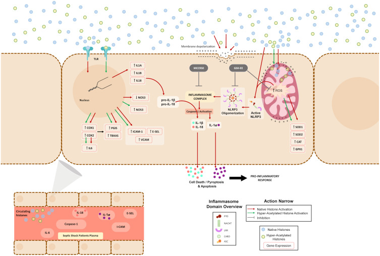
Circulating extracellular histones acquire relevance as cytotoxic mediators in sepsis. Extracellular histones act as damage-associated molecular patterns (DAMPs), which induce oxidative stress and NLRP3 inflammasome activation. Inflammasome mediates pyroptosis, a programmed cell death mechanism that produces inflammation. Despite evidence for inflammasome activation in immune cells during sepsis, it was unknown whether extracellular histones can produce endothelial inflammasomes activation.
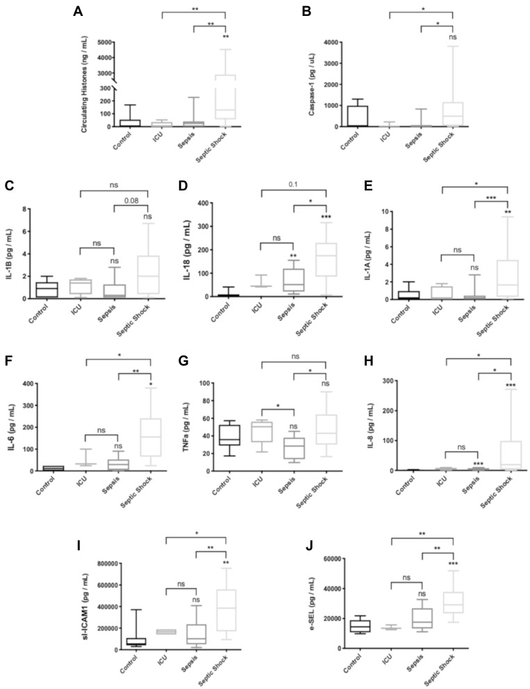
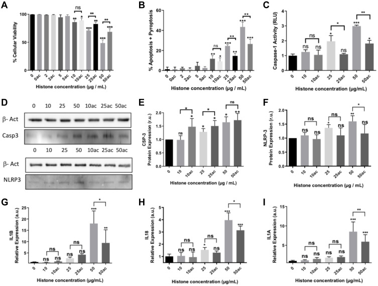
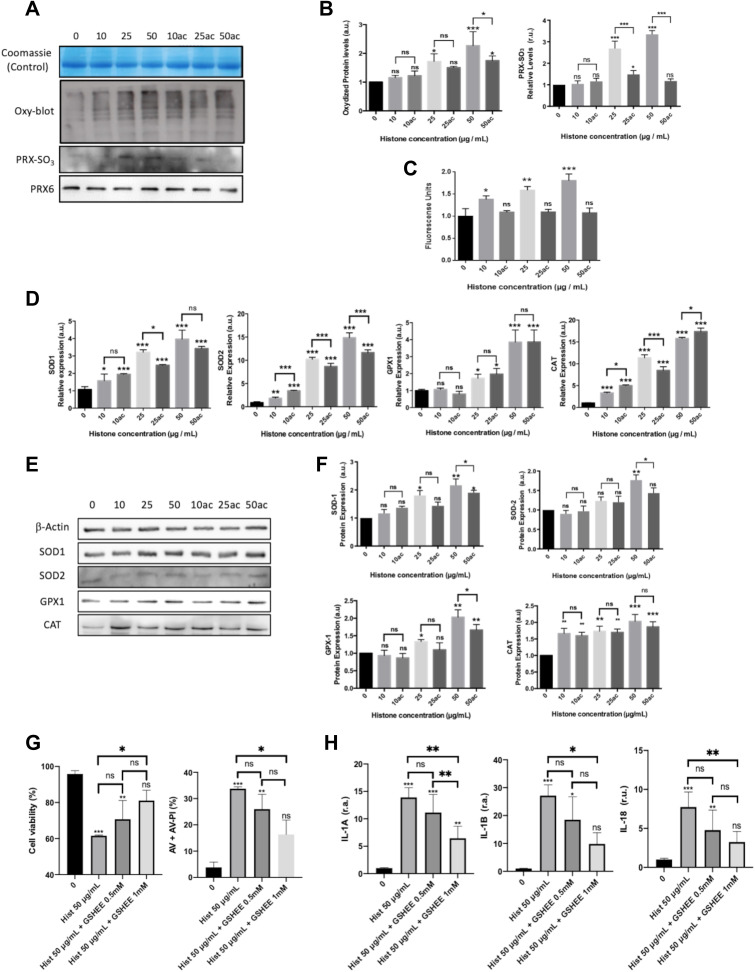
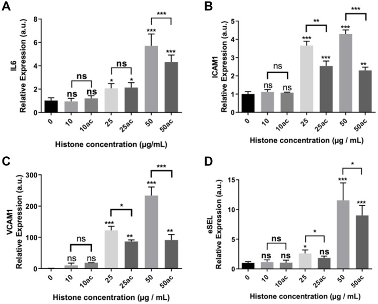
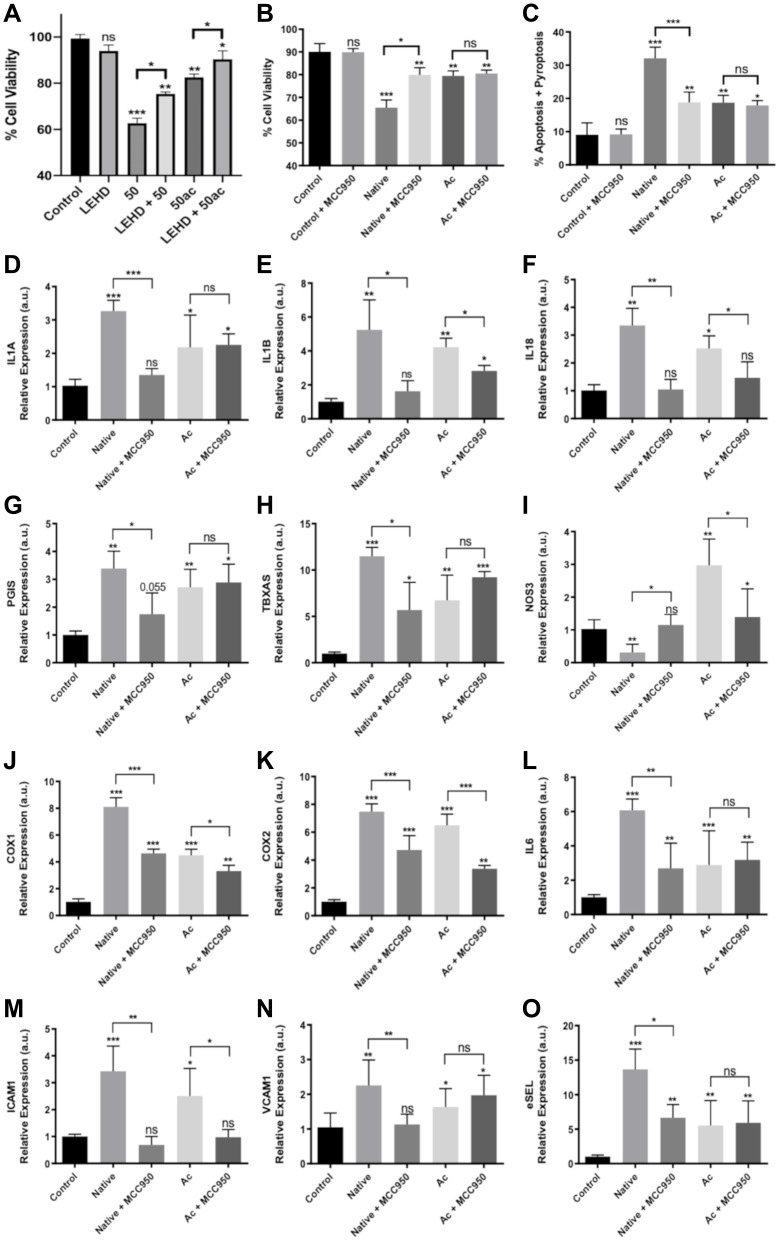
We used human umbilical vein endothelial cells (HUVEC) to explore the activation of pyroptosis, endothelial function and inflammation by extracellular histones. We evaluated pyroptosis by flow cytometry, caspase-1 activity assay, and gene and protein expression analysis by RT-qPCR and Western blot, respectively. The upstream molecular responses involved in pyroptosis activation by extracellular histones were validated by means of using antioxidant glutathione ethyl ester and NLRP3 inflammasome inhibitors. Finally, using mass spectrometry, we measured circulating histones in blood from critically-ill patients and demonstrated that circulating histone levels correlated with the expression of pyroptosis-related cytokines, the release of endothelial adhesion factors and septic shock severity.
We found that extracellular histones mediate the activation of NLRP3 inflammasome and pyroptosis in endothelial cells by contributing to endothelial dysfunction and the dysregulation of the immune response mediated by endothelium. Likewise, we demonstrated how the hyperacetylation of extracellular histones or the use of antioxidants decreased pyroptosis. In addition, we showed that pyroptosis is a feasible process occurring in septic shock patients.
Circulating histone levels correlated with the expression of pro-inflammatory and pyroptosis-related cytokines, the release of endothelial adhesion factors and septic shock severity. We propose to block histone-mediated pyroptosis as a feasible therapeutic strategy in sepsis.
循环细胞外组蛋白在脓毒症中作为细胞毒性介质具有重要意义。细胞外组蛋白作为损伤相关分子模式(DAMPs),可诱导氧化应激和NLRP3炎性小体激活。炎性小体介导细胞焦亡,这是一种产生炎症的程序性细胞死亡机制。尽管有证据表明脓毒症期间免疫细胞中炎性小体被激活,但细胞外组蛋白是否能激活内皮细胞炎性小体尚不清楚。
我们使用人脐静脉内皮细胞(HUVEC)来探究细胞外组蛋白对细胞焦亡、内皮功能和炎症的激活作用。我们分别通过流式细胞术、半胱天冬酶-1活性测定以及RT-qPCR和蛋白质印迹法进行基因和蛋白质表达分析来评估细胞焦亡。通过使用抗氧化剂谷胱甘肽乙酯和NLRP3炎性小体抑制剂验证了细胞外组蛋白激活细胞焦亡所涉及的上游分子反应。最后,我们使用质谱法测量了危重症患者血液中的循环组蛋白,并证明循环组蛋白水平与细胞焦亡相关细胞因子的表达、内皮黏附因子的释放以及脓毒性休克的严重程度相关。
我们发现细胞外组蛋白通过导致内皮功能障碍和内皮介导的免疫反应失调,介导内皮细胞中NLRP3炎性小体的激活和细胞焦亡。同样,我们证明了细胞外组蛋白的高乙酰化或使用抗氧化剂可减少细胞焦亡。此外,我们表明细胞焦亡是脓毒性休克患者中发生的一个可行过程。
循环组蛋白水平与促炎和细胞焦亡相关细胞因子的表达、内皮黏附因子的释放以及脓毒性休克的严重程度相关。我们提出阻断组蛋白介导的细胞焦亡作为脓毒症中一种可行的治疗策略。