Al Rasheed Raghad, Adhi Mohammad Idrees, Alowedi Sarah Abdullah, Albdah Bayan, Aldebasi Tariq, Hazzazi Mohammad A
Department of Ophthalmology, King Abdulaziz Medical City, National Guard Health Affairs, PO Box 22490, Riyadh, 11426, Saudi Arabia.
King Saud Bin Abdulaziz University for Health Sciences, Riyadh, Saudi Arabia.
Int J Retina Vitreous. 2022 Aug 2;8(1):53. doi: 10.1186/s40942-022-00402-3.
Few challenges are faced with the introduction of anti-VEGF agents as a modality of treatment for retinopathy of prematurity. The clinical behavior and time course of regression post injection differ compared to post laser ablation. This study aims to evaluate the long-term peripheral retinal vascularization outcome of Ranibizumab intravitreal injections monotherapy in the treatment of retinopathy of prematurity.
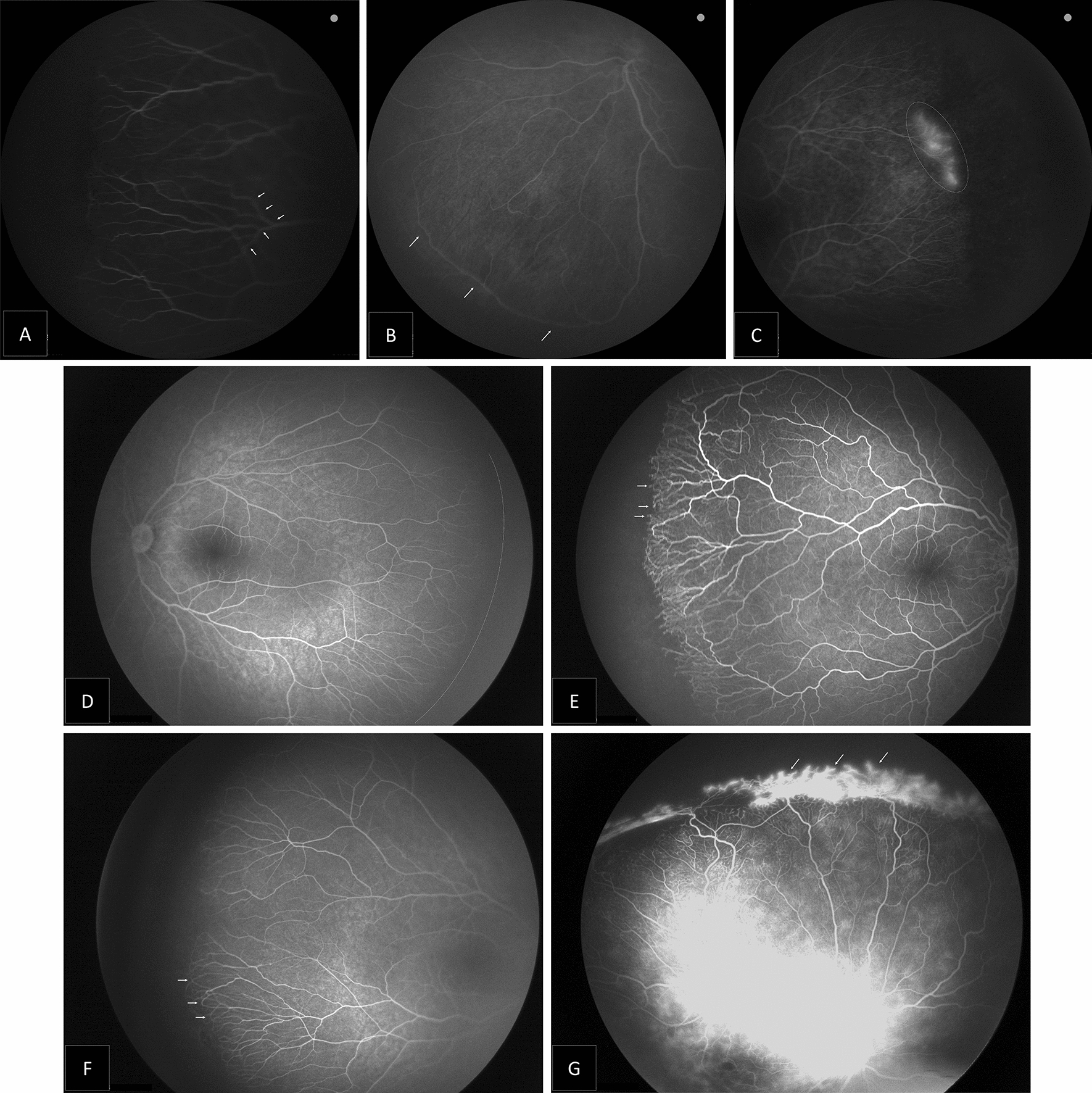
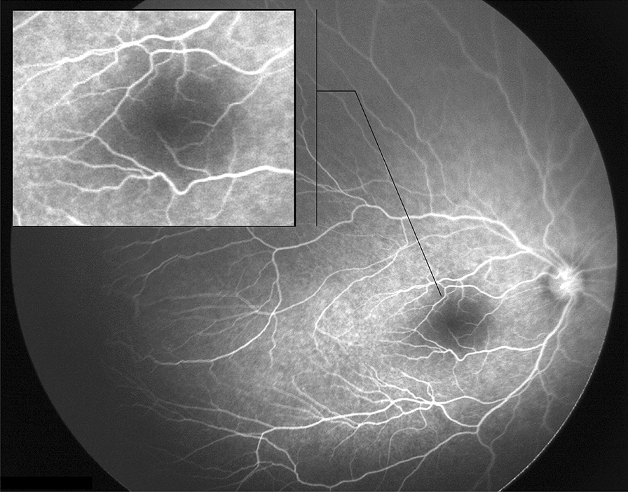
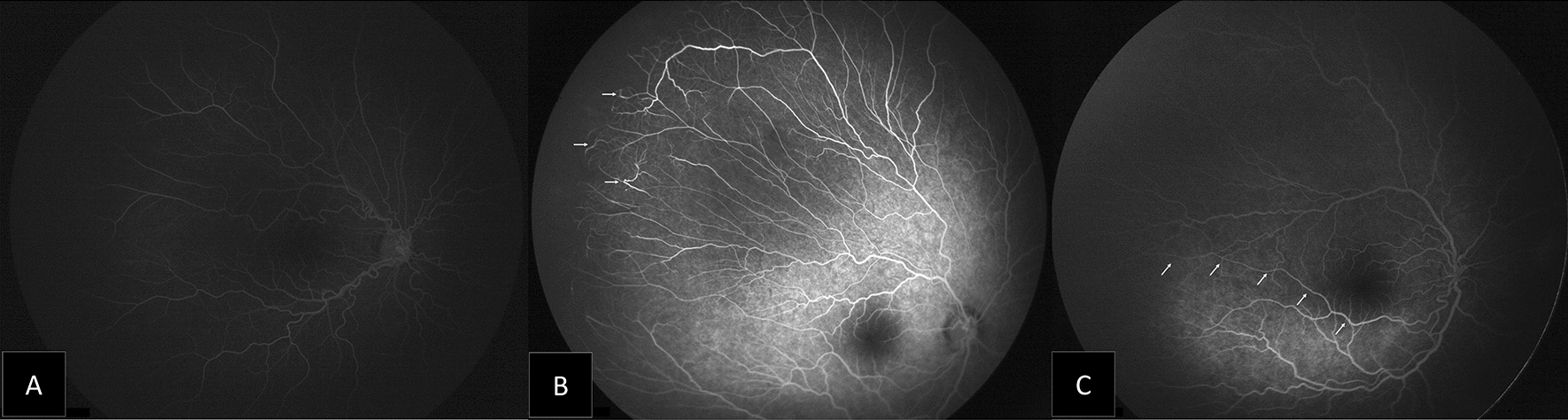
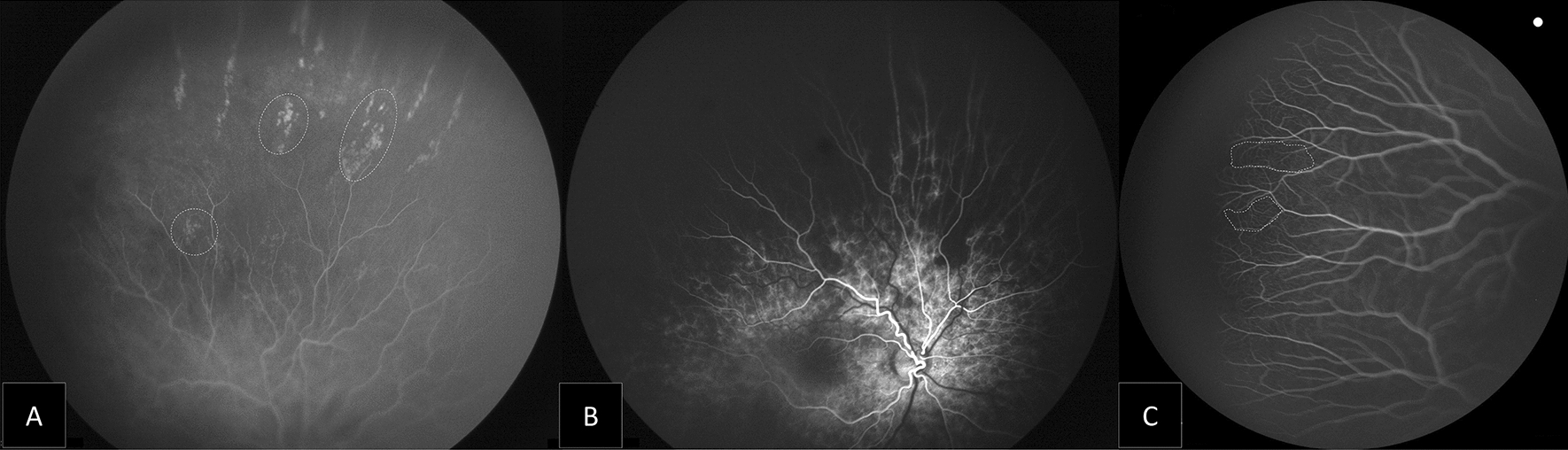
Hospital-based quasi-experimental study. Include ROP patients who received intravitreal ranibizumab (IVR), as primary treatment for type 1 ROP. Patients were examined under general anaesthesia to ensure documentation of all junctions of vascular and avascular zones. Images were taken by RetCam III, Phoenix ICON and fluorescein angiography was performed to describe vascular behaviors.
The mean gestational age was 24.67 weeks and the mean postmenstrual age at the time of intravitreal ranibizumab treatment was 36.3 weeks. Fluorescein angiography was performed at 155-288 weeks; most eyes showed two disk diameters of avascular peripheral retina. Only eyes with original aggressive ROP who required a second injection (six eyes) showed extensive peripheral avascular retina reaching zone I (13.64%). Neovascularization was evident in five eyes (11.36%), all with an original aggressive ROP and received multiple injections.
Ranibizumab treated babies with incomplete retinal vascularization require close and long-term follow-up visits to assess post injection vascular behavior. Peripheral retinal avascular zone of more than two-disc diameters was present in most of the patients evidenced by fluorescein angiography. Babies with initial diagnosis of aggressive ROP are more likely to have persistent peripheral neovascularization.
将抗血管内皮生长因子(VEGF)药物作为早产儿视网膜病变的一种治疗方式面临的挑战较少。与激光消融术后相比,注射后消退的临床行为和时间进程有所不同。本研究旨在评估玻璃体内注射雷珠单抗单药治疗早产儿视网膜病变的长期周边视网膜血管化结果。
基于医院的准实验研究。纳入接受玻璃体内注射雷珠单抗(IVR)作为1型早产儿视网膜病变主要治疗方法的患者。在全身麻醉下对患者进行检查,以确保记录血管和无血管区域的所有交界处。使用RetCam III拍摄图像,进行Phoenix ICON检查,并进行荧光素血管造影以描述血管行为。
平均胎龄为24.67周,玻璃体内注射雷珠单抗治疗时的平均月经后年龄为36.3周。在155 - 288周进行荧光素血管造影;大多数眼睛显示周边有无血管视网膜,直径为两个视盘。只有最初患有侵袭性早产儿视网膜病变且需要第二次注射的眼睛(6只眼)显示广泛的周边无血管视网膜延伸至I区(13.64%)。5只眼(11.36%)出现新生血管,所有这些眼睛最初均患有侵袭性早产儿视网膜病变并接受了多次注射。
雷珠单抗治疗的视网膜血管化不完全的婴儿需要密切和长期的随访,以评估注射后的血管行为。荧光素血管造影显示大多数患者的周边视网膜无血管区直径超过两个视盘。最初诊断为侵袭性早产儿视网膜病变的婴儿更有可能出现持续性周边新生血管。