Division of Infectious Diseases and HIV Medicine, Department of Medicine, Case Western Reserve University, Cleveland, OH, USA.
Department of Pediatrics, Case Western Reserve University, Cleveland, OH, USA.
Diabetologia. 2022 Dec;65(12):2157-2171. doi: 10.1007/s00125-022-05775-6. Epub 2022 Aug 3.
AIMS/HYPOTHESIS: CD40 expressed in Müller cells is a central driver of diabetic retinopathy. CD40 causes phospholipase Cγ1 (PLCγ1)-dependent ATP release in Müller cells followed by purinergic receptor (P2X)-dependent production of proinflammatory cytokines in myeloid cells. In the diabetic retina, CD40 and P2X upregulate a broad range of inflammatory molecules that promote development of diabetic retinopathy. The molecular event downstream of CD40 that activates the PLCγ1-ATP-P2X-proinflammatory cytokine cascade and promotes development of diabetic retinopathy is unknown. We hypothesise that disruption of the CD40-driven molecular events that trigger this cascade prevents/treats diabetic retinopathy in mice.
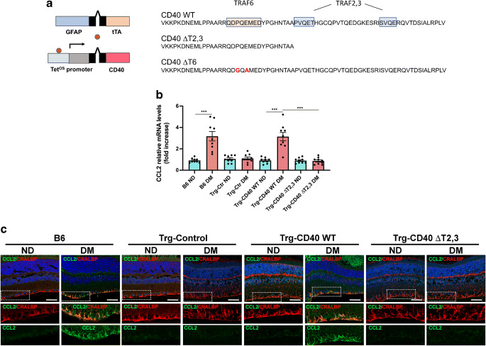
B6 and transgenic mice with Müller cell-restricted expression of wild-type (WT) CD40 or CD40 with mutations in TNF receptor-associated factor (TRAF) binding sites were made diabetic using streptozotocin. Leucostasis was assessed using FITC-conjugated concanavalin A. Histopathology was examined in the retinal vasculature. Expression of inflammatory molecules and phospho-Tyr783 PLCγ1 (p-PLCγ1) were assessed using real-time PCR, immunoblot and/or immunohistochemistry. Release of ATP and cytokines were measured by ATP bioluminescence and ELISA, respectively.
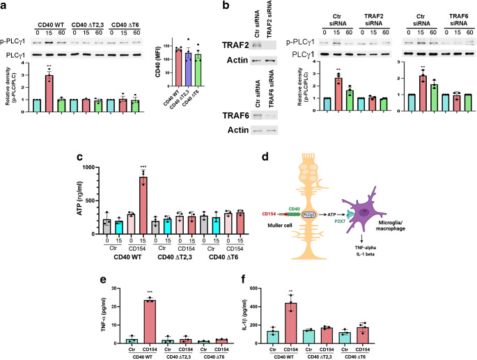
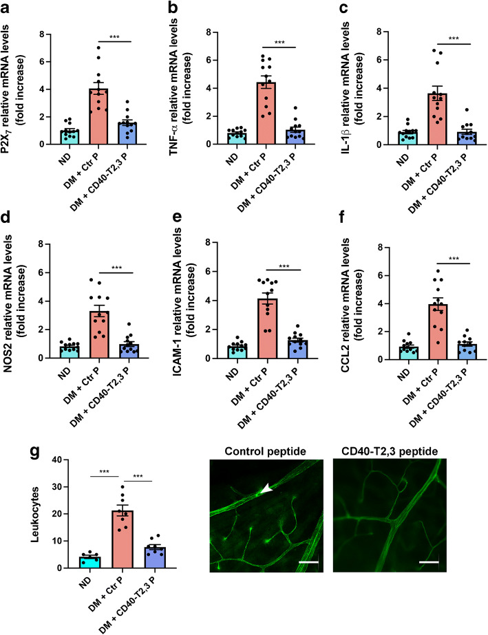
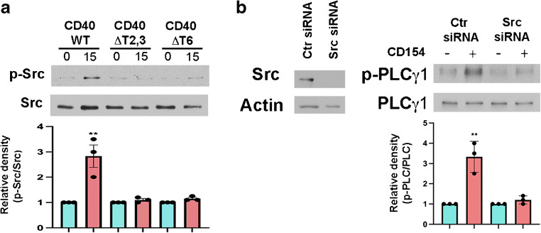
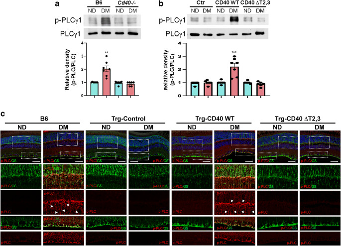
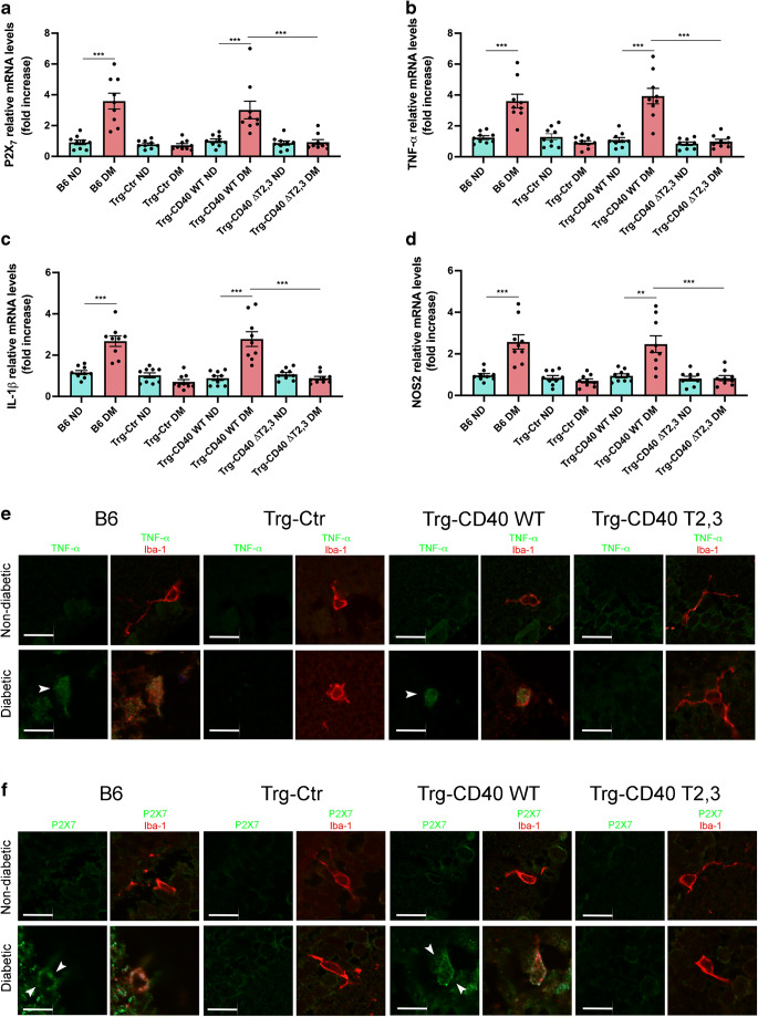
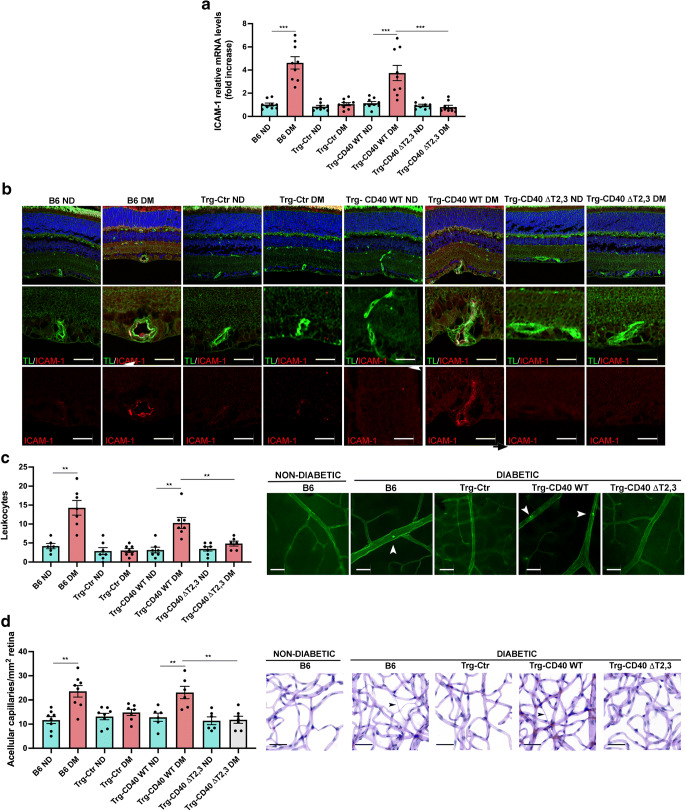
Human Müller cells with CD40 ΔT2,3 (lacks TRAF2,3 binding sites) were unable to phosphorylate PLCγ1 and release ATP in response to CD40 ligation, and could not induce TNF-α/IL-1β secretion in bystander myeloid cells. CD40-TRAF signalling acted via Src to induce PLCγ1 phosphorylation. Diabetic mice in which WT CD40 in Müller cells was replaced by CD40 ΔT2,3 failed to exhibit phosphorylation of PLCγ1 in these cells and upregulate P2X and TNF-α in microglia/macrophages. P2x (also known as P2rx7), Tnf-α (also known as Tnf), Il-1β (also known as Il1b), Nos2, Icam-1 (also known as Icam1) and Ccl2 mRNA were not increased in these mice and the mice did not develop retinal leucostasis and capillary degeneration. Diabetic B6 mice treated intravitreally with a cell-permeable peptide that disrupts CD40-TRAF2,3 signalling did not exhibit either upregulation of P2X and inflammatory molecules in the retina or leucostasis.
CONCLUSIONS/INTERPRETATION: CD40-TRAF2,3 signalling activated the CD40-PLCγ1-ATP-P2X-proinflammatory cytokine pathway. Src functioned as a link between CD40-TRAF2,3 and PLCγ1. Replacing WT CD40 with CD40 ΔT2,3 impaired activation of PLCγ1 in Müller cells, upregulation of P2X in microglia/macrophages, upregulation of a broad range of inflammatory molecules in the diabetic retina and the development of diabetic retinopathy. Administration of a peptide that disrupts CD40-TRAF2,3 signalling reduced retinal expression of inflammatory molecules and reduced leucostasis in diabetic mice, supporting the therapeutic potential of pharmacological inhibition of CD40-TRAF2,3 in diabetic retinopathy.
目的/假设:在 Müller 细胞中表达的 CD40 是糖尿病性视网膜病变的核心驱动因素。CD40 导致 PLCγ1(磷脂酶 Cγ1)依赖性 ATP 在 Müller 细胞中的释放,随后在髓样细胞中通过嘌呤能受体(P2X)依赖性产生促炎细胞因子。在糖尿病视网膜中,CD40 和 P2X 上调了广泛的促炎分子,促进了糖尿病性视网膜病变的发展。激活 PLCγ1-ATP-P2X-促炎细胞因子级联反应并促进糖尿病性视网膜病变发展的 CD40 下游的分子事件尚不清楚。我们假设破坏触发该级联反应的 CD40 驱动的分子事件可预防/治疗小鼠的糖尿病性视网膜病变。
使用链脲佐菌素使 B6 和具有 Müller 细胞中野生型(WT)CD40 或具有 TNF 受体相关因子(TRAF)结合位点突变的 CD40 过表达的转基因小鼠发生糖尿病。使用 FITC 缀合的刀豆球蛋白 A 评估白细胞增多症。在视网膜血管中检查组织病理学。使用实时 PCR、免疫印迹和/或免疫组织化学评估炎症分子和磷酸化 Tyr783 PLCγ1(p-PLCγ1)的表达。通过 ATP 生物发光和 ELISA 分别测量 ATP 和细胞因子的释放。
缺乏 TRAF2,3 结合位点的人 Müller 细胞中的 CD40 ΔT2,3 无法响应 CD40 连接而磷酸化 PLCγ1 并释放 ATP,并且不能诱导旁观者髓样细胞中 TNF-α/IL-1β 的分泌。CD40-TRAF 信号通过Src 诱导 PLCγ1 磷酸化。用 CD40 ΔT2,3 替换 Müller 细胞中 WT CD40 的糖尿病小鼠未能在这些细胞中观察到 PLCγ1 的磷酸化,并在上皮细胞/巨噬细胞中上调 P2X 和 TNF-α。这些小鼠中 P2x(也称为 P2rx7)、Tnf-α(也称为 Tnf)、Il-1β(也称为 Il1b)、Nos2、Icam-1(也称为 Icam1)和 Ccl2 mRNA 没有增加,并且这些小鼠没有发生视网膜白细胞增多症和毛细血管变性。用一种可渗透细胞的肽在玻璃体内治疗糖尿病 B6 小鼠,该肽破坏 CD40-TRAF2,3 信号传导,不会导致视网膜中 P2X 和炎症分子的上调或白细胞增多症。
结论/解释:CD40-TRAF2,3 信号激活了 CD40-PLCγ1-ATP-P2X-促炎细胞因子途径。Src 作为 CD40-TRAF2,3 和 PLCγ1 之间的联系。用 CD40 ΔT2,3 替代 WT CD40 会损害 Müller 细胞中 PLCγ1 的激活,微胶质细胞/巨噬细胞中 P2X 的上调,糖尿病视网膜中广泛的炎症分子的上调以及糖尿病性视网膜病变的发展。施用破坏 CD40-TRAF2,3 信号的肽可减少糖尿病小鼠视网膜中炎症分子的表达并减少白细胞增多症,这支持了在糖尿病性视网膜病变中药理学抑制 CD40-TRAF2,3 的治疗潜力。