Sriamornrattanakul Kitiporn, Akharathammachote Nasaeng, Chonhenchob Areeporn, Mongkolratnan Atithep, Niljianskul Nattawut, Phoominaonin I-Sorn, Ariyaprakai Chanon, Wongsuriyanan Somkiat
Department of Surgery, Faculty of Medicine Vajira Hospital, Navamindradhiraj University, Bangkok, Thailand.
Department of Health Technology, Faculty of Science and Health Technology, Navamindradhiraj University, Bangkok, Thailand.
Surg Neurol Int. 2022 Jul 15;13:304. doi: 10.25259/SNI_346_2022. eCollection 2022.
The third segment of the vertebral artery (V3) is vulnerable during far lateral and retrosigmoid approaches. Although the suboccipital triangle (SOT) is a useful anatomical landmark, the relationship between V3 and the muscles forming the triangle is not well-described. We aimed to demonstrate the relationship between the V3, surrounding muscles, and SOT in clinical cases.
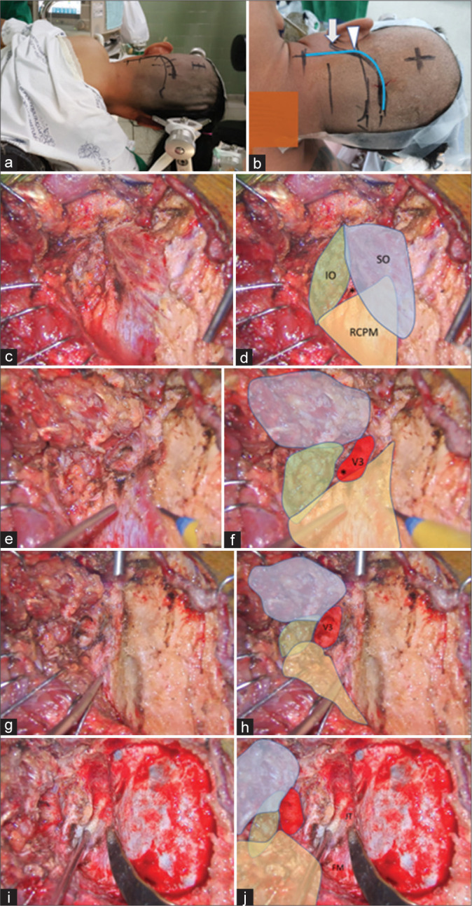
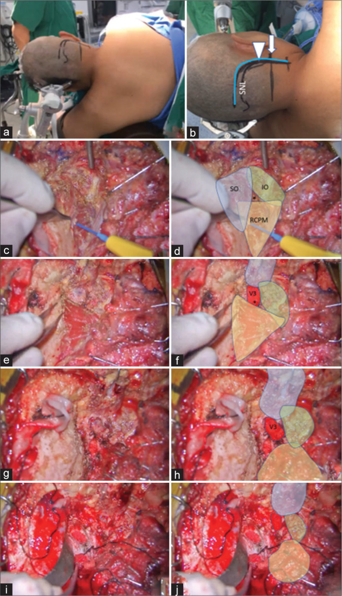
Operative videos of patients with the vertebral artery (VA) and posterior inferior cerebellar artery (PICA) aneurysms treated with occipital artery-PICA bypass through the far lateral approach were examined. Videos from January 2015 to October 2021 were retrospectively reviewed to determine anatomy of the V3 and the SOT.
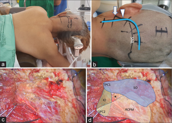
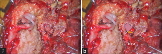
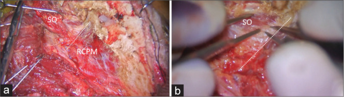
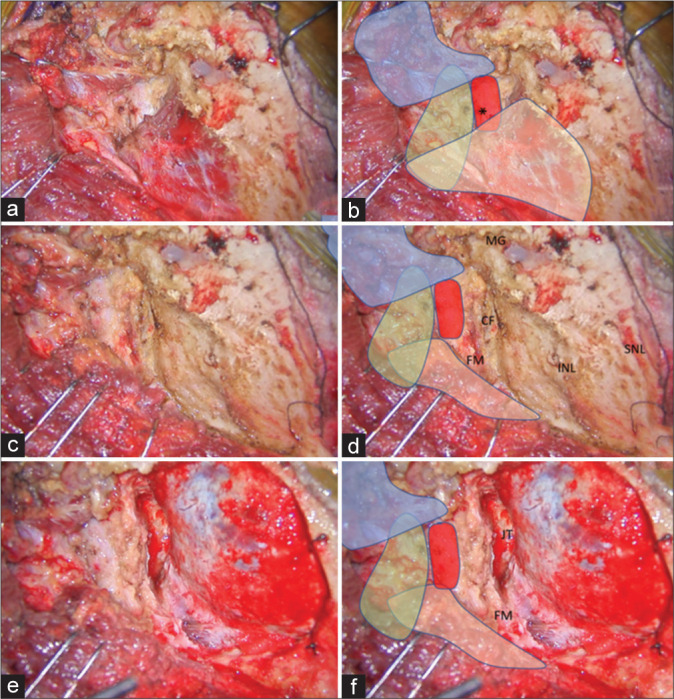
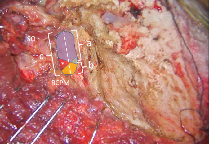
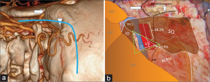
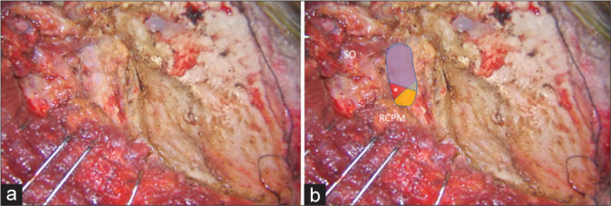
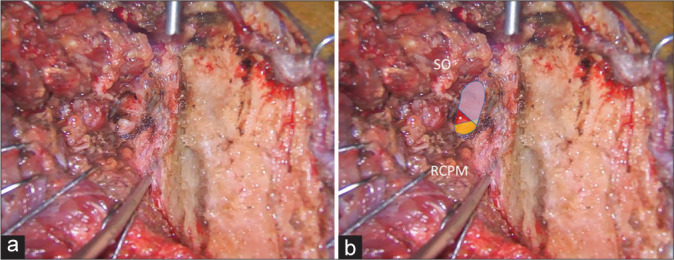
Fourteen patients were included in this study. The ipsilateral V3 was identified without injury in all patients using the bipolar cutting technique. The lateral 68.2% of the horizontal V3 segment, including the V3 bulge, was covered by the inferomedial part of the superior oblique muscle (SO). The medial 23.9% was covered by the inferolateral part of the rectus capitis posterior major muscle. The inferomedial part of the horizontal V3 segment is located within the SOT.
Most of the V3, including the V3 bulge, were located beneath the SO and the inferomedial part of V3 located within the SOT. Elevation of the SO should be performed carefully using the bipolar cutting technique to avoid injury to the V3. To the best of our knowledge, this is the first description of the V3 relative to the SOT in the clinical setting.
在远外侧入路和乙状窦后入路手术中,椎动脉第三段(V3)易受损伤。尽管枕下三角(SOT)是一个有用的解剖标志,但V3与构成该三角的肌肉之间的关系尚未得到充分描述。我们旨在通过临床病例展示V3、周围肌肉和SOT之间的关系。
检查经远外侧入路行枕动脉-小脑后下动脉(PICA)搭桥术治疗椎动脉(VA)和小脑后下动脉瘤患者的手术视频。回顾性分析2015年1月至2021年10月的视频,以确定V3和SOT的解剖结构。
本研究纳入14例患者。所有患者均采用双极电切技术,在未损伤同侧V3的情况下识别出该血管。水平V3段的外侧68.2%,包括V3膨大部,被上斜肌(SO)的下内侧部分覆盖。内侧23.9%被头后大直肌的下外侧部分覆盖。水平V3段的下内侧部分位于SOT内。
大部分V3,包括V3膨大部,位于SO下方,V3的下内侧部分位于SOT内。应使用双极电切技术小心抬起SO,以避免损伤V3。据我们所知,这是在临床环境中首次描述V3与SOT的关系。