Liu Haitao, Hong Xupeng, Xi Ji, Menne Stephan, Hu Jianming, Wang Joseph Che-Yen
Department of Microbiology and Immunology, The Pennsylvania State University College of Medicine, Hershey, PA 17033, USA.
Department of Microbiology and Immunology, Georgetown University Medical Center, Washington DC 20007, USA.
Sci Adv. 2022 Aug 5;8(31):eabo4184. doi: 10.1126/sciadv.abo4184.
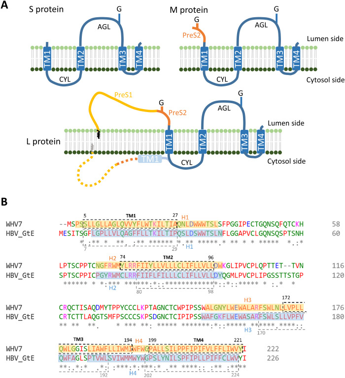
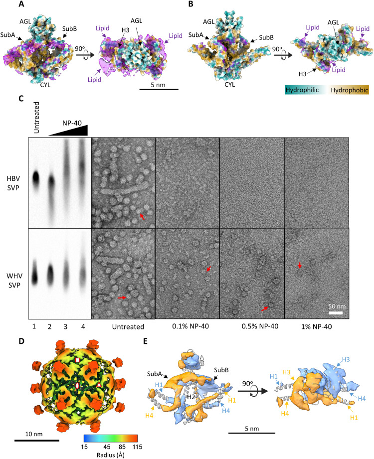
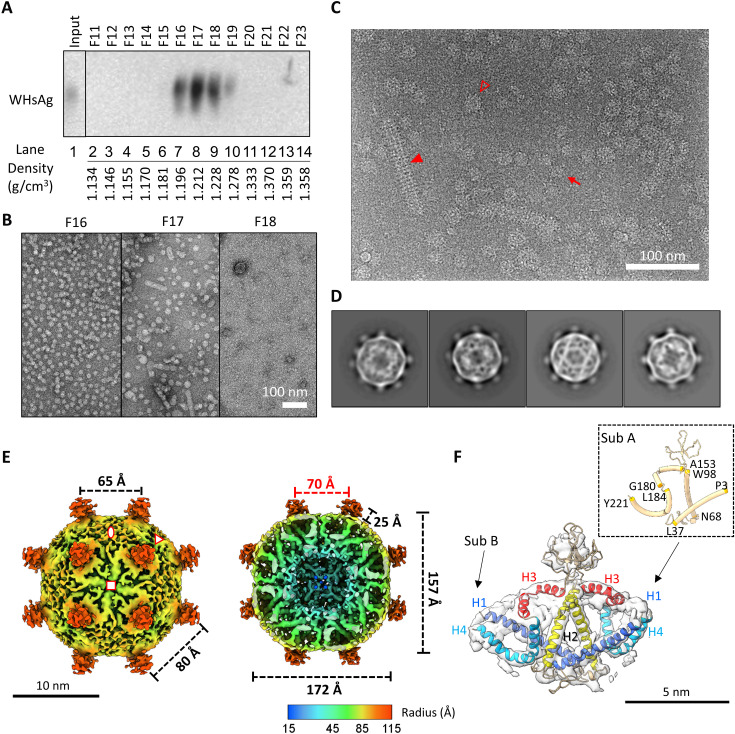
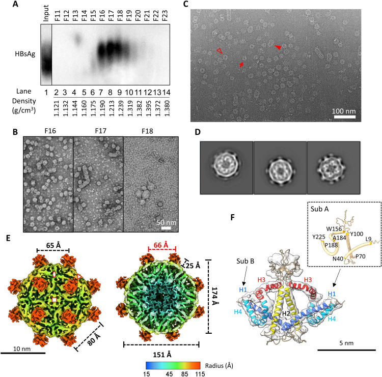
The loss of detectable hepatitis B surface antigen (HBsAg) is considered a functional cure in chronic hepatitis B. Naturally, HBsAg can be incorporated into the virion envelope or assembled into subviral particles (SVPs) with lipid from host cells. Until now, there has been no detailed structure of HBsAg, and the published SVP structures are controversial. Here, we report the first subnanometer-resolution structures of spherical SVP from hepatitis B virus (HBV) and the related woodchuck hepatitis virus (WHV) determined by cryo-electron microscopy in combination with AlphaFold2 prediction. Both structures showed unique rhombicuboctahedral symmetry with 24 protruding spikes comprising dimer of small HBsAg with four helical domains. The lipid moiety in the SVP is organized in a noncanonical lipid patch instead of a lipid bilayer, which can accommodate the exposed hydrophobic surface and modulate particle stability. Together, these findings advance our knowledge of viral membrane organization and the structures of HBV and WHV spherical SVPs.
可检测到的乙型肝炎表面抗原(HBsAg)的消失被认为是慢性乙型肝炎的功能性治愈。自然情况下,HBsAg可整合到病毒粒子包膜中,或与宿主细胞的脂质组装成亚病毒颗粒(SVP)。到目前为止,尚未有HBsAg的详细结构报道,且已发表的SVP结构存在争议。在此,我们报告了通过冷冻电子显微镜结合AlphaFold2预测确定的来自乙型肝炎病毒(HBV)和相关土拨鼠肝炎病毒(WHV)的球形SVP的首个亚纳米分辨率结构。两种结构均显示出独特的菱形二十面体对称性,有24个突出的刺突,由具有四个螺旋结构域的小HBsAg二聚体组成。SVP中的脂质部分以非典型脂质斑块而非脂质双层的形式组织,可容纳暴露的疏水表面并调节颗粒稳定性。这些发现共同推进了我们对病毒膜组织以及HBV和WHV球形SVP结构的认识。