Department of Gerontological Nursing/Wound Care Management, Graduate School of Medicine, The University of Tokyo, Tokyo, Japan.
Japan Society for the Promotion of Science, Tokyo, Japan.
Int Wound J. 2023 Mar;20(3):648-658. doi: 10.1111/iwj.13906. Epub 2022 Aug 6.
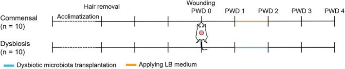
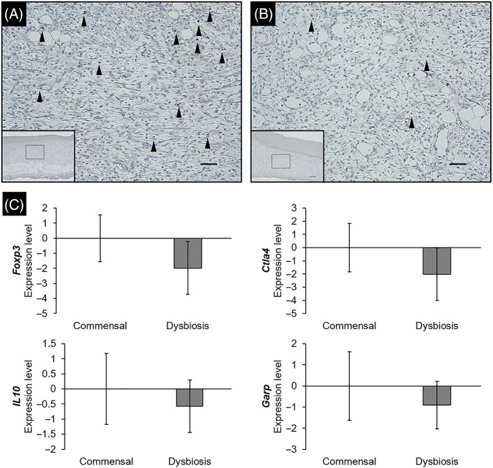
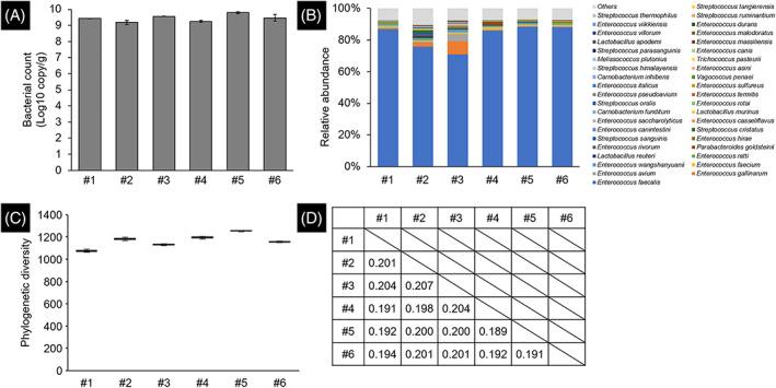
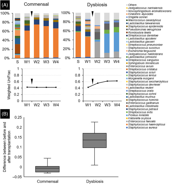
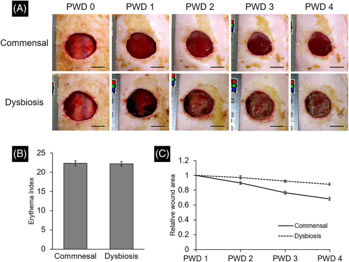
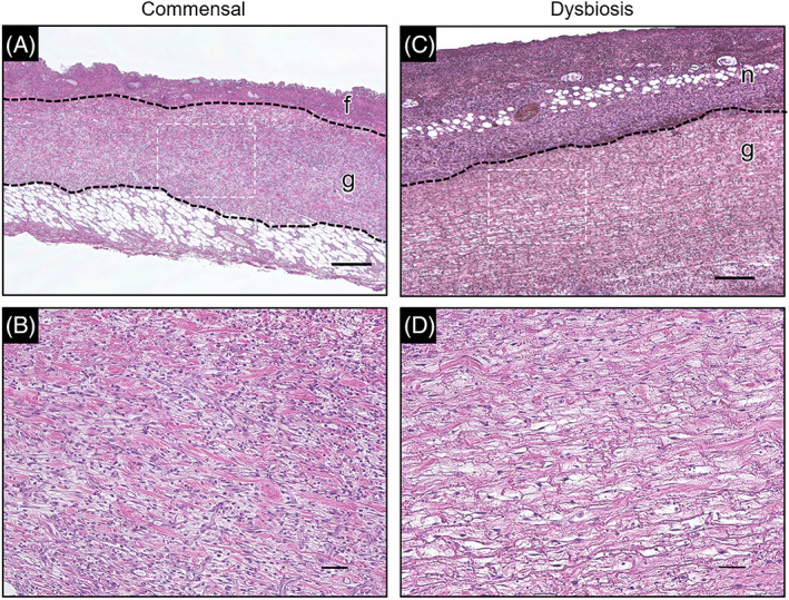
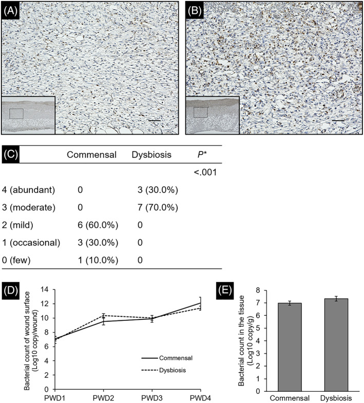
In critically colonised wounds, many of the signs of infection are often absent, and delayed healing may be the only clinical sign. The prevention of critical colonisation is important, but its pathophysiology has not yet been elucidated. We have previously reported that dysbiotic microbiota dissimilar to the peri-wound skin microbiota may develop in critically colonised wounds. To investigate the role of dysbiotic microbiota, this study aimed to develop a critically colonised wound model by transplantation of dysbiotic microbiota. To transplant microbiota, a bacterial solution (dysbiosis group) or with Luria-Bertani medium (commensal group) was inoculated to full-thickness wounds of rats. The bacterial solution was prepared by anaerobically culturing bacteria from donor rats on an artificial dermis in Luria-Bertani medium for 72 hours. As a result, the degree of the change in the microbial similarity between pre- and post-transplantation of microbiota was significantly higher in the dysbiosis group (P < .001). No signs of infection were observed in any rat in either group. The wound area in the dysbiosis group was significantly larger (P < .001), and there was a significant infiltration of neutrophils (P < .001). All rats of the dysbiosis group represented the clinical features of critically colonised wounds. Furthermore, there were significantly fewer regulatory T cells in the wounds of the dysbiosis group. This is the first study to develop a novel animal model that represents the clinical features of critically colonised wounds and will be useful in investigating the pathogenesis of critical colonisation via regulatory T cells.
在严重定植的伤口中,许多感染迹象通常不存在,延迟愈合可能是唯一的临床迹象。预防严重定植很重要,但它的病理生理学尚未阐明。我们之前报道过,在严重定植的伤口中可能会出现与伤口周围皮肤微生物群不同的失调微生物群。为了研究失调微生物群的作用,本研究旨在通过移植失调微生物群来建立严重定植的伤口模型。为了移植微生物群,将细菌溶液(失调组)或含 Luria-Bertani 培养基(共生组)接种到大鼠全层伤口中。细菌溶液是通过在 Luria-Bertani 培养基中的人工真皮上厌氧培养供体大鼠的细菌 72 小时制备的。结果,微生物群移植前后微生物相似性变化的程度在失调组中显著更高(P <.001)。两组大鼠均未出现感染迹象。失调组的伤口面积明显更大(P <.001),并且中性粒细胞浸润明显(P <.001)。所有失调组的大鼠均表现出严重定植伤口的临床特征。此外,失调组伤口中的调节性 T 细胞明显减少。这是第一项开发代表严重定植伤口临床特征的新型动物模型的研究,将有助于通过调节性 T 细胞研究严重定植的发病机制。