Department of Surgery, Stomatology, Pathology and Radiology, Bauru School of Dentistry, University of São Paulo, Bauru, Brazil.
OMFS-IMPATH Research Group, Department of Imaging and Pathology, Faculty of Medicine, KU Leuven and Oral and Maxillofacial Surgery, University Hospitals Leuven, Leuven, Belgium.
Clin Exp Dent Res. 2022 Dec;8(6):1487-1495. doi: 10.1002/cre2.643. Epub 2022 Aug 7.
The aim of this study is to investigate the long-term effects on jaw and femur bone induced by oncologic doses of zoledronic acid in a young rat model.
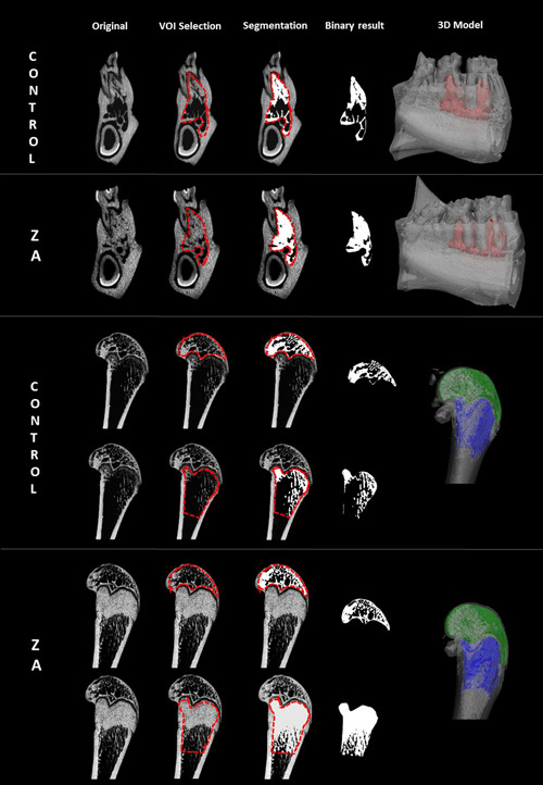
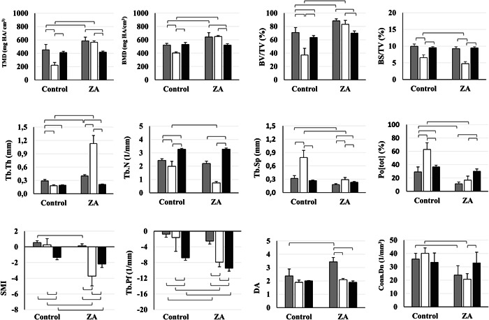
Six 12-week-old male Wistar rats received zoledronic acid (0.6 mg/kg) and six control rats received saline solution in the same volume. Compounds were administered intraperitoneally in five doses every 28 days. Euthanasia was performed 150 days after therapy onset. After animal sacrifice, their mandibles and femurs were scanned ex vivo using a high-resolution (14 μm) micro-computed tomography. Morphometric bone parameters were calculated using CT-Analyzer (Bruker, Belgium) between the first and second mandibular molars and in the distal femur metaphysis and epiphysis.
The treatment group as compared to the controls showed a significantly (p < .05) increased bone quantity (↑BV/TV, ↓Po[Tot], ↑Tb.Th), bone density (↑TMD, ↑BMD), and osteosclerosis of the trabecular bone (↓Tb.Sp, ↓Conn.Dn, ↓Tb.Pf, ↓SMI) in all anatomical sites. Bone remodeling suppression due to zoledronic acid treatment was more pronounced (p < .05) in the femoral metaphysis relative to the mandible and epiphysis. The exploratory linear discriminant analysis showed that for the mandible, it was mainly the bone quantity-related morphometric indices (BV/TV and Tb.Th), while for the femoral epiphysis and metaphysis, it was bone structure-related (Tb.Pf and Tb.N), which are of primary importance to study the treatment effect.
High doses of bisphosphonates can differently affect the bone quantity, density, and structure in long bones and jawbones. In the metaphysis, bone changes were primarily concentrated in the region of the growth plate. Future studies may consider the use of bone morphometric indices to evaluate the effect of bisphosphonates.
本研究旨在探讨唑来膦酸在年轻大鼠模型中对颌骨和股骨的长期影响。
6 只 12 周龄雄性 Wistar 大鼠接受唑来膦酸(0.6mg/kg),6 只对照大鼠接受相同体积的生理盐水。化合物通过腹腔内注射每 28 天 5 次给予。治疗开始后 150 天进行安乐死。动物牺牲后,使用高分辨率(14μm)微计算机断层扫描对其下颌骨和股骨进行离体扫描。使用 CT-Analyzer(Bruker,比利时)在下颌第一和第二磨牙之间以及股骨远端干骺端和骨骺处计算形态计量骨参数。
与对照组相比,治疗组在所有解剖部位均表现出显著(p<0.05)增加的骨量(↑BV/TV,↓Po[Tot],↑Tb.Th)、骨密度(↑TMD,↑BMD)和小梁骨的骨硬化(↓Tb.Sp,↓Conn.Dn,↓Tb.Pf,↓SMI)。由于唑来膦酸治疗导致的骨重塑抑制在股骨干骺端比下颌骨和骨骺更明显(p<0.05)。探索性线性判别分析表明,对于下颌骨,主要是与骨量相关的形态计量学指标(BV/TV 和 Tb.Th),而对于股骨骨骺和干骺端,主要是与骨结构相关的(Tb.Pf 和 Tb.N),这些指标对于研究治疗效果至关重要。
高剂量的双膦酸盐可以不同地影响长骨和颌骨的骨量、密度和结构。在干骺端,骨变化主要集中在生长板区域。未来的研究可能会考虑使用骨形态计量学指标来评估双膦酸盐的作用。