Hsu Pei-Yu, Tsai Ming-Tzu, Wang Shun-Ping, Chen Ying-Ju, Wu Jay, Hsu Jui-Ting
Department of Biomedical Imaging and Radiological Science, China Medical University, Taichung, 404, Taiwan.
Department of Biomedical Engineering, Hungkuang University, Taichung, Taiwan, 433, ROC.
PLoS One. 2016 Apr 29;11(4):e0154367. doi: 10.1371/journal.pone.0154367. eCollection 2016.
This study used microcomputed tomography (micro-CT) to evaluate the effects of ovariectomy on the trabecular bone microarchitecture and cortical bone morphology in the femoral neck and mandible of female rats.
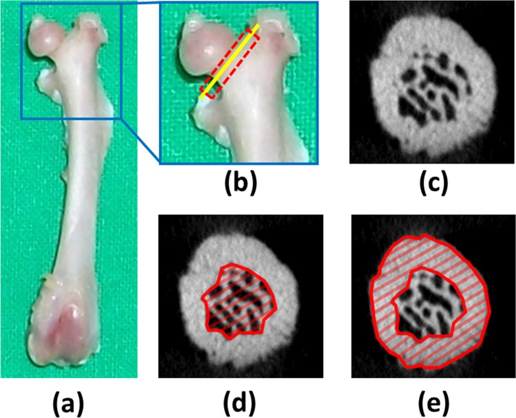
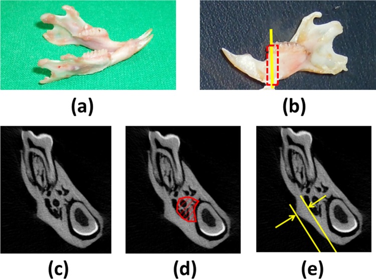
Twelve female Wister rats were divided into two groups: the control and ovariectomized groups. The rats in the ovariectomized group received ovariectomy at 8 weeks of age; all the rats were sacrificed at 20 weeks of age, and their mandibles and femurs were removed and scanned using micro-CT. Four microstructural trabecular bone parameters were measured for the region below the first mandibular molar and the femoral neck region: bone volume fraction (BV/TV), trabecular thickness (TbTh), trabecular separation (TbSp), and trabecular number (TbN). In addition, four cortical bone parameters were measured for the femoral neck region: total cross-sectional area (TtAr), cortical area (CtAr), cortical bone area fraction (CtAr/TtAr), and cortical thickness (CtTh). The CtTh at the masseteric ridge was used to assess the cortical bone morphology in the mandible. The trabecular bone microarchitecture and cortical bone morphology in the femoral necks and mandibles of the control group were compared with those of the ovariectomized group. Furthermore, Spearman's correlation (rs) was conducted to analyze the correlation between the osteoporosis conditions of the mandible and femoral neck.
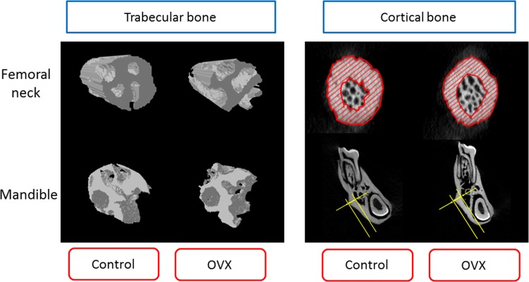
Regarding the trabecular bone microarchitectural parameters, the BV/TV of the trabecular bone microarchitecture in the femoral necks of the control group (61.199±11.288%, median ± interquartile range) was significantly greater than that of the ovariectomized group (40.329±5.153%). Similarly, the BV/TV of the trabecular bone microarchitecture in the mandibles of the control group (51.704±6.253%) was significantly greater than that of the ovariectomized group (38.486±9.111%). Furthermore, the TbSp of the femoral necks in the ovariectomized group (0.185±0.066 mm) was significantly greater than that in the control group (0.130±0.026mm). Similarly, the TbSp of the mandibles in the ovariectomized group (0.322±0.047mm) was significantly greater than that in the control group (0.285±0.041mm). However, the TbTh and TbN trends for the mandibles and femoral necks were inconsistent between the control and ovariectomized groups. Regarding the cortical bone morphology parameters, the TtAr of the femoral necks in the ovariectomized group was significantly smaller than that in the control group. There was no significant difference in the TtAr, CtAr, or CtTh of the femoral necks between the control and ovariectomized groups, and no significant difference in the CtTh of the mandibles between the control and ovariectomized groups. Moreover, the BV/TV and TbSp of the mandibles were highly correlated with those of the femurs (rs = 0.874 and rs = 0.755 for BV/TV and TbSp, respectively). Nevertheless, the TbTh, TbN, and CtTh of the mandibles were not correlated with those of the femoral necks.
After the rats were ovariectomized, osteoporosis of the trabecular bone microarchitecture occurred in their femurs and mandibles; however, ovariectomy did not influence the cortical bone morphology. In addition, the parametric values of the trabecular bone microarchitecture in the femoral necks were highly correlated with those of the trabecular bone microarchitecture in the mandibles.
本研究采用显微计算机断层扫描(micro-CT)评估卵巢切除对雌性大鼠股骨颈和下颌骨小梁骨微结构及皮质骨形态的影响。
将12只雌性Wister大鼠分为两组:对照组和卵巢切除组。卵巢切除组的大鼠在8周龄时接受卵巢切除术;所有大鼠在20周龄时处死,取出其下颌骨和股骨并使用micro-CT进行扫描。测量下颌第一磨牙下方区域和股骨颈区域的四个小梁骨微结构参数:骨体积分数(BV/TV)、小梁厚度(TbTh)、小梁间距(TbSp)和小梁数量(TbN)。此外,测量股骨颈区域的四个皮质骨参数:总横截面积(TtAr)、皮质面积(CtAr)、皮质骨面积分数(CtAr/TtAr)和皮质厚度(CtTh)。咬肌嵴处的CtTh用于评估下颌骨的皮质骨形态。比较对照组和卵巢切除组大鼠股骨颈和下颌骨的小梁骨微结构及皮质骨形态。此外,进行Spearman相关性分析(rs)以分析下颌骨和股骨颈骨质疏松状况之间的相关性。
关于小梁骨微结构参数,对照组股骨颈小梁骨微结构的BV/TV(61.199±11.288%,中位数±四分位间距)显著大于卵巢切除组(40.329±5.153%)。同样,对照组下颌骨小梁骨微结构的BV/TV(51.704±6.253%)显著大于卵巢切除组(38.486±9.111%)。此外,卵巢切除组股骨颈的TbSp(0.185±0.066mm)显著大于对照组(0.130±0.026mm)。同样,卵巢切除组下颌骨的TbSp(0.322±0.047mm)显著大于对照组(0.285±0.041mm)。然而,对照组和卵巢切除组之间下颌骨和股骨颈的TbTh和TbN趋势不一致。关于皮质骨形态参数,卵巢切除组股骨颈的TtAr显著小于对照组。对照组和卵巢切除组股骨颈的TtAr、CtAr或CtTh之间无显著差异,对照组和卵巢切除组下颌骨的CtTh之间也无显著差异。此外,下颌骨的BV/TV和TbSp与股骨的BV/TV和TbSp高度相关(BV/TV和TbSp的rs分别为0.874和0.755)。然而,下颌骨的TbTh、TbN和CtTh与股骨颈的TbTh、TbN和CtTh不相关。
大鼠卵巢切除后,其股骨和下颌骨出现小梁骨微结构骨质疏松;然而,卵巢切除未影响皮质骨形态。此外,股骨颈小梁骨微结构的参数值与下颌骨小梁骨微结构的参数值高度相关。