• 文献检索
  • 文档翻译
  • 深度研究
  • 学术资讯
  • Suppr Zotero 插件Zotero 插件
  • 邀请有礼
  • 套餐&价格
  • 历史记录
应用&插件
Suppr Zotero 插件Zotero 插件浏览器插件Mac 客户端Windows 客户端微信小程序
定价
高级版会员购买积分包购买API积分包
服务
文献检索文档翻译深度研究API 文档MCP 服务
关于我们
关于 Suppr公司介绍联系我们用户协议隐私条款
关注我们

Suppr 超能文献

核心技术专利:CN118964589B侵权必究
粤ICP备2023148730 号-1Suppr @ 2026

文献检索

告别复杂PubMed语法,用中文像聊天一样搜索,搜遍4000万医学文献。AI智能推荐,让科研检索更轻松。

立即免费搜索

文件翻译

保留排版,准确专业,支持PDF/Word/PPT等文件格式,支持 12+语言互译。

免费翻译文档

深度研究

AI帮你快速写综述,25分钟生成高质量综述,智能提取关键信息,辅助科研写作。

立即免费体验

磁共振成像在颅内出血和脑梗死中的影像特征的诊断价值。

Diagnostic Value of Image Features of Magnetic Resonance Imaging in Intracranial Hemorrhage and Cerebral Infarction.

机构信息

Department of Radiology, The Second Affiliated Hospital of Hainan Medical University, Haikou 570000, Hainan, China.

Department of Radiology, Hainan Danzhou People's Hospital, Danzhou 571700, Hainan, China.

出版信息

Contrast Media Mol Imaging. 2022 Jul 12;2022:6495568. doi: 10.1155/2022/6495568. eCollection 2022.

DOI:10.1155/2022/6495568
PMID:35935302
原文链接:https://pmc.ncbi.nlm.nih.gov/articles/PMC9296345/
Abstract

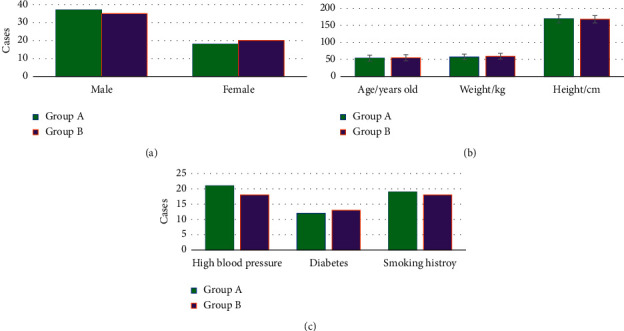
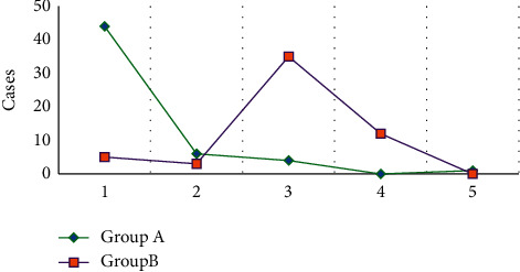
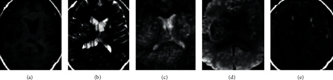
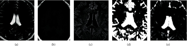
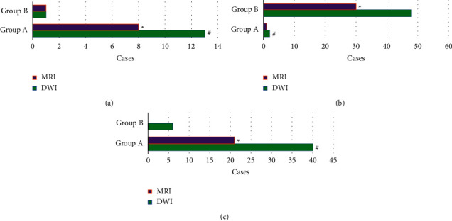
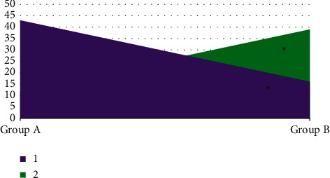
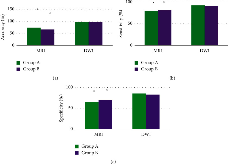
This study aimed to investigate the differential diagnosis value of routine magnetic resonance imaging (MRI) and magnetic resonance diffusion-weighted imaging (DWI) in hyperacute intracranial hemorrhage (HICH) and hyperacute cerebral infarction (HCI). Fifty-five patients with HICH were set as group A, and 55 patients with HCI were selected as group B. All the patients underwent routine MRI and DWI examinations. The morphological distribution and signal characteristics (low, high, or mixed) of the lesions in the two groups were recorded. The diagnostic accuracy, sensitivity, and specificity of routine MRI and DWI were compared for distinguishing HICH and HCI. The results suggested that the lesions in patients with HICH were mainly manifested as mixed signals (40 cases), while those in patients with HCI showed high signals (48 cases). HICH occurred in the basal ganglia in 44 cases, in the brain stem in 6 cases, in the cerebellum in 4 cases, in the cerebral cortex in 0 cases, and in the corpus callosum in 1 case. HCI occurred in the basal ganglia area, brain stem, cerebellum, cerebral cortex, and corpus callosum in 5, 3, 35, 12, and 0 cases, respectively. The diagnostic accuracy, specificity, and sensitivity of DWI for HICH and HCI were significantly higher than those of routine MRI ( < 0.05). It was indicated that compared with routine MRI, DWI was more effective in the diagnosis of HICH and HCI, with clearer and more accurate images and better diagnostic performance.

摘要

本研究旨在探讨常规磁共振成像(MRI)和磁共振弥散加权成像(DWI)在超急性颅内出血(HICH)和超急性脑梗死(HCI)中的鉴别诊断价值。将 55 例 HICH 患者设为 A 组,55 例 HCI 患者设为 B 组。所有患者均行常规 MRI 和 DWI 检查,记录两组患者病灶的形态分布和信号特征(低、高或混杂)。比较常规 MRI 和 DWI 鉴别 HICH 和 HCI 的诊断准确性、灵敏度和特异度。结果表明,HICH 患者病灶主要表现为混杂信号(40 例),HCI 患者病灶表现为高信号(48 例)。HICH 发生于基底节 44 例,脑干 6 例,小脑 4 例,大脑皮质 0 例,胼胝体 1 例。HCI 发生于基底节区、脑干、小脑、大脑皮质和胼胝体分别为 5、3、35、12 和 0 例。DWI 对 HICH 和 HCI 的诊断准确性、特异性和灵敏度均明显高于常规 MRI(<0.05)。表明与常规 MRI 相比,DWI 对 HICH 和 HCI 的诊断更有效,图像更清晰、准确,诊断效能更好。

https://cdn.ncbi.nlm.nih.gov/pmc/blobs/3cd5/9296345/605495c0465e/CMMI2022-6495568.007.jpg
https://cdn.ncbi.nlm.nih.gov/pmc/blobs/3cd5/9296345/d20b1c8acc4e/CMMI2022-6495568.001.jpg
https://cdn.ncbi.nlm.nih.gov/pmc/blobs/3cd5/9296345/b46a38d39d78/CMMI2022-6495568.002.jpg
https://cdn.ncbi.nlm.nih.gov/pmc/blobs/3cd5/9296345/4734c4234063/CMMI2022-6495568.003.jpg
https://cdn.ncbi.nlm.nih.gov/pmc/blobs/3cd5/9296345/2a12d5165a67/CMMI2022-6495568.004.jpg
https://cdn.ncbi.nlm.nih.gov/pmc/blobs/3cd5/9296345/81e5a1ff96f8/CMMI2022-6495568.005.jpg
https://cdn.ncbi.nlm.nih.gov/pmc/blobs/3cd5/9296345/d86dff6c8011/CMMI2022-6495568.006.jpg
https://cdn.ncbi.nlm.nih.gov/pmc/blobs/3cd5/9296345/605495c0465e/CMMI2022-6495568.007.jpg

相似文献

1
Diagnostic Value of Image Features of Magnetic Resonance Imaging in Intracranial Hemorrhage and Cerebral Infarction.磁共振成像在颅内出血和脑梗死中的影像特征的诊断价值。
Contrast Media Mol Imaging. 2022 Jul 12;2022:6495568. doi: 10.1155/2022/6495568. eCollection 2022.
2
[Early diagnostic significance and dynamic pattern of DWI compared with conventional MRI in newborns with neonatal cerebral infarction].[新生儿脑梗死患儿中磁共振扩散加权成像(DWI)与传统磁共振成像(MRI)对比的早期诊断意义及动态变化模式]
Zhonghua Er Ke Za Zhi. 2007 May;45(5):360-4.
3
[Application of diffusion-weighted and perfusion magnetic resonance imaging in definition of the ischemic penumbra in hyperacute cerebral infarction].[弥散加权和灌注磁共振成像在超急性脑梗死缺血半暗带定义中的应用]
Zhonghua Yi Xue Za Zhi. 2003 Jun 10;83(11):952-7.
4
[Early assessment of severe hypoxic-ischemic encephalopathy in neonates by diffusion-weighted magnetic resonance imaging techniques and its significance].[磁共振扩散加权成像技术对新生儿重度缺氧缺血性脑病的早期评估及其意义]
Zhonghua Er Ke Za Zhi. 2007 Nov;45(11):843-7.
5
Proton magnetic resonance spectroscopy and diffusion-weighted imaging in intracranial cystic mass lesions.颅内囊性肿块病变的质子磁共振波谱和扩散加权成像
Surg Neurol. 2007;68 Suppl 1:S25-36. doi: 10.1016/j.surneu.2007.07.080.
6
[Values of diffusion-weighted magnetic resonance imaging in the early detection of periventricular leukomalacia in preterm infants].[扩散加权磁共振成像在早产儿脑室周围白质软化早期检测中的价值]
Zhonghua Er Ke Za Zhi. 2008 May;46(5):354-8.
7
Callosal Diffusion-Restriction in Patients with Intracranial Hemorrhage.胼胝体弥散受限患者。
J Stroke Cerebrovasc Dis. 2021 Sep;30(9):105951. doi: 10.1016/j.jstrokecerebrovasdis.2021.105951. Epub 2021 Jul 21.
8
Significance of clinical-diffusion mismatch in hyperacute cerebral infarction.超急性期脑梗死中临床-弥散不匹配的意义。
J Stroke Cerebrovasc Dis. 2011 Jan-Feb;20(1):62-67. doi: 10.1016/j.jstrokecerebrovasdis.2009.11.002. Epub 2010 Jun 17.
9
Diagnostic Value of Magnetic Resonance Diffusion-Weighted Imaging Combined with Routine Scanning in Body Tumors.磁共振扩散加权成像联合常规扫描对体部肿瘤的诊断价值。
Contrast Media Mol Imaging. 2022 Jul 21;2022:5799815. doi: 10.1155/2022/5799815. eCollection 2022.
10
Contribution of diffusion-weighted magnetic resonance imaging in the characterization of hepatocellular carcinomas and dysplastic nodules in cirrhotic liver.扩散加权磁共振成像在肝硬化肝脏中肝细胞癌和发育异常结节特征描述中的作用
J Comput Assist Tomogr. 2010 Jul;34(4):506-12. doi: 10.1097/RCT.0b013e3181da3671.

本文引用的文献

1
The value of diffusion weighted imaging-alberta stroke program early CT score in predicting stroke-associated pneumonia in patients with acute cerebral infarction: a retrospective study.弥散加权成像-阿尔伯塔卒中项目早期 CT 评分在预测急性脑梗死患者卒中相关性肺炎中的价值:一项回顾性研究。
PeerJ. 2022 Jan 19;10:e12789. doi: 10.7717/peerj.12789. eCollection 2022.
2
Ischemic stroke in oral anticoagulated patients with atrial fibrillation.口服抗凝药物治疗的心房颤动患者的缺血性脑卒中。
Acta Neurol Scand. 2022 Mar;145(3):288-296. doi: 10.1111/ane.13552. Epub 2021 Nov 12.
3
Image Features of Magnetic Resonance Angiography under Deep Learning in Exploring the Effect of Comprehensive Rehabilitation Nursing on the Neurological Function Recovery of Patients with Acute Stroke.
基于深度学习的磁共振血管成像的图像特征在探索综合康复护理对急性脑卒中患者神经功能恢复的影响中的应用。
Contrast Media Mol Imaging. 2021 Sep 10;2021:1197728. doi: 10.1155/2021/1197728. eCollection 2021.
4
Fuzzy System Based Medical Image Processing for Brain Disease Prediction.基于模糊系统的用于脑部疾病预测的医学图像处理
Front Neurosci. 2021 Jul 30;15:714318. doi: 10.3389/fnins.2021.714318. eCollection 2021.
5
Risk factors and outcomes of hospitalized stroke patients in Lusaka, Zambia.赞比亚卢萨卡住院脑卒中患者的风险因素和结局。
J Neurol Sci. 2021 May 15;424:117404. doi: 10.1016/j.jns.2021.117404. Epub 2021 Mar 18.
6
[Embolic stroke of undetermined source (ESUS) - Classification of a new stroke entity].[不明来源栓塞性卒中(ESUS)——一种新的卒中类型的分类]
Dtsch Med Wochenschr. 2021 Mar;146(6):403-409. doi: 10.1055/a-1309-8701. Epub 2021 Mar 18.
7
Which Imaging Approach Should Be Used for Stroke of Unknown Time of Onset?不明时间起病脑卒中患者应选择哪种影像学方法?
Stroke. 2021 Jan;52(1):373-380. doi: 10.1161/STROKEAHA.120.032020. Epub 2020 Dec 11.
8
Trousseau Syndrome Related Cerebral Infarction: Clinical Manifestations, Laboratory Findings and Radiological Features.特鲁索综合征相关脑梗死:临床表现、实验室检查及影像学特征。
J Stroke Cerebrovasc Dis. 2020 Sep;29(9):104891. doi: 10.1016/j.jstrokecerebrovasdis.2020.104891. Epub 2020 Jun 30.
9
Advanced Machine-Learning Methods for Brain-Computer Interfacing.高级机器学习方法在脑机接口中的应用。
IEEE/ACM Trans Comput Biol Bioinform. 2021 Sep-Oct;18(5):1688-1698. doi: 10.1109/TCBB.2020.3010014. Epub 2021 Oct 7.
10
[Not Available].[无可用内容]。
Rev Med Interne. 2020 Jul;41(7):469-474. doi: 10.1016/j.revmed.2020.04.018. Epub 2020 Jul 24.