Appana Dalavi Pandurang, Prabhu Ashwini, M Sajida, Chatterjee Kaushik, Venkatesan Jayachandran
Biomaterials Research Laboratory, Yenepoya Research Centre, Yenepoya (Deemed to be University), Mangalore 575018, India.
Department of Materials Engineering, Indian Institute of Science, Bangalore 560012, India.
ACS Omega. 2022 Jul 21;7(30):26092-26106. doi: 10.1021/acsomega.2c00995. eCollection 2022 Aug 2.
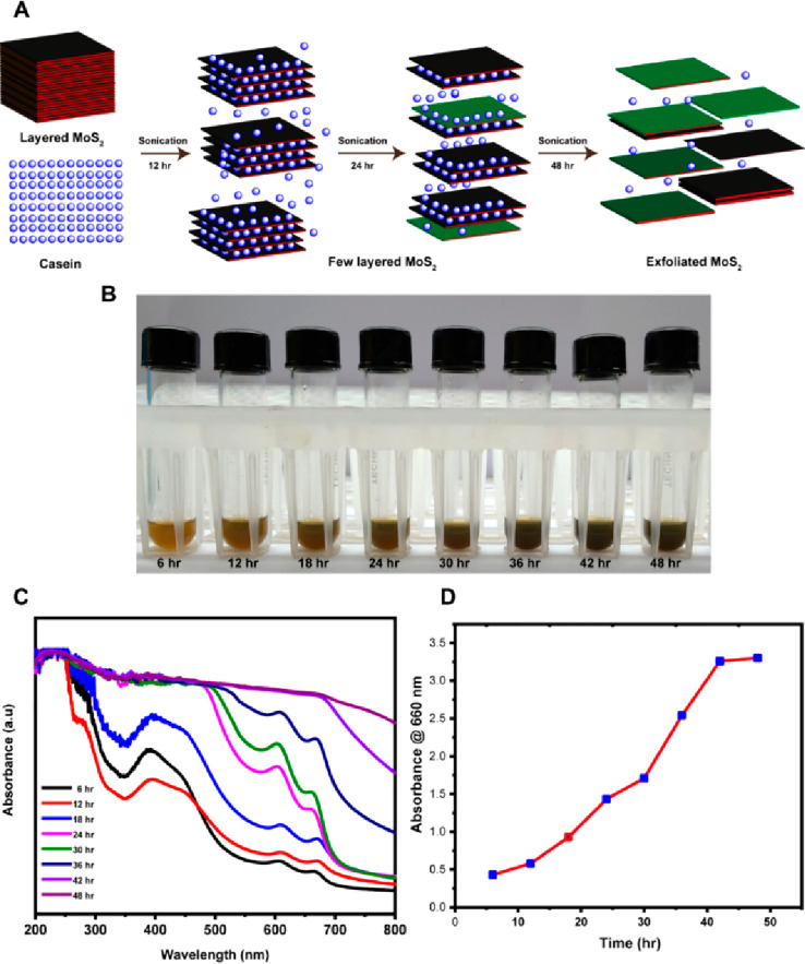
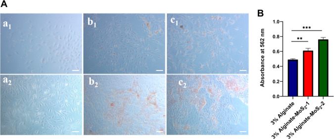
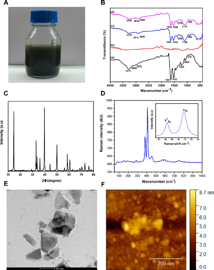
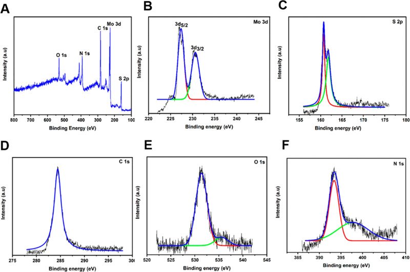
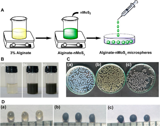
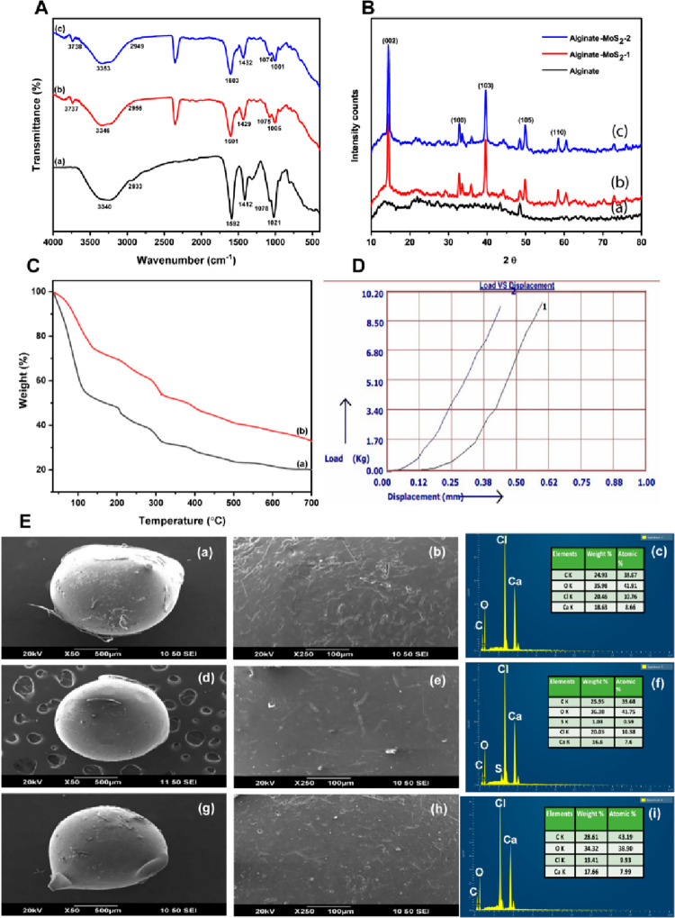
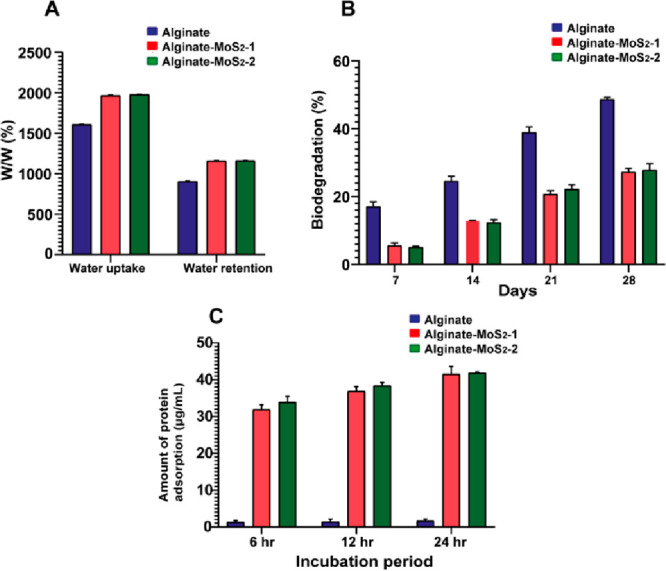
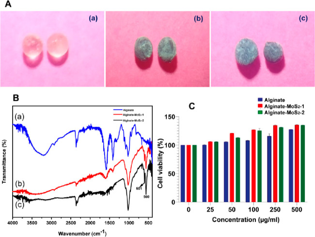
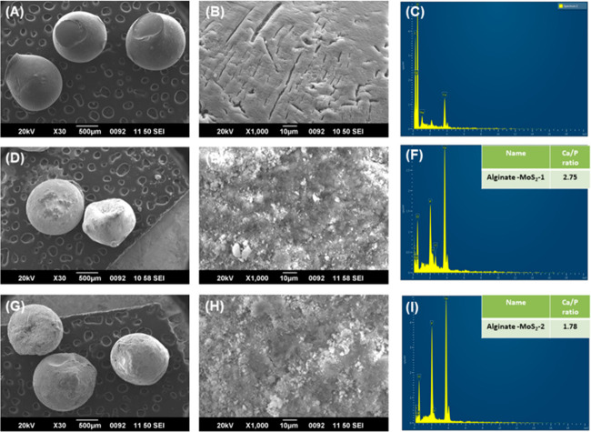
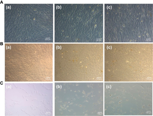
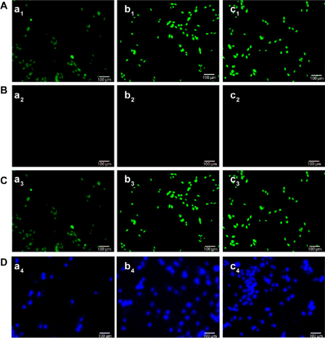
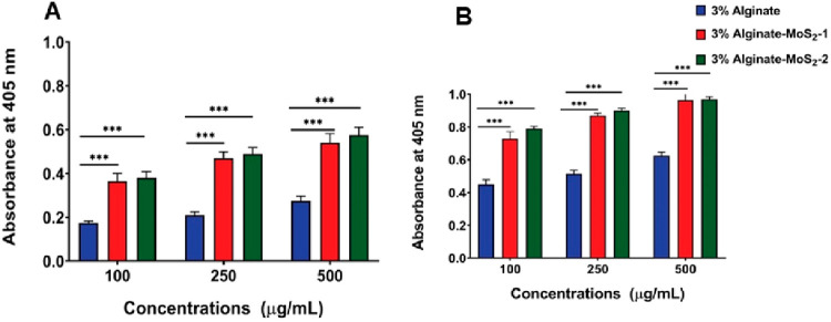
Defects and disorders of the bone due to disease, trauma, or abnormalities substantially affect a person's life quality. Research in bone tissue engineering is motivated to address these clinical needs. The present study demonstrates casein-mediated liquid exfoliation of molybdenum disulfide (MoS) and its coupling with alginate to create microspheres to engineer bone graft substitutes. Casein-exfoliated nano-MoS was chemically characterized using different analytical techniques. The UV-visible spectrum of nano-MoS-2 displayed strong absorption peaks at 610 and 668 nm. In addition, the XPS spectra confirmed the presence of the molybdenum (Mo, 3d), sulfur (S, 2p), carbon (C, 1s), oxygen (O, 1s), and nitrogen (N, 1s) elements. The exfoliated MoS nanosheets were biocompatible with the MG-63, MC3T3-E1, and C2C12 cells at 250 μg/mL concentration. Further, microspheres were created using alginate, and they were characterized physiochemically and biologically. Stereomicroscopic images showed that the microspheres were spherical with an average diameter of 1 ± 0.2 mm. The dispersion of MoS in the alginate matrix was uniform. The alginate-MoS microspheres promoted apatite formation in the SBF (simulated body fluid) solution. Moreover, the alginate-MoS was biocompatible with MG-63 cells and promoted cell proliferation. Higher alkaline phosphatase activity and mineralization were observed on the alginate-MoS with the MG-63 cells. Hence, the developed alginate-MoS microsphere could be a potential candidate for a bone graft substitute.
由疾病、创伤或异常引起的骨骼缺陷和疾病会严重影响人的生活质量。骨组织工程的研究旨在满足这些临床需求。本研究展示了酪蛋白介导的二硫化钼(MoS)液体剥离及其与海藻酸盐的偶联,以制备微球用于构建骨移植替代物。使用不同的分析技术对酪蛋白剥离的纳米MoS进行了化学表征。纳米MoS-2的紫外可见光谱在610和668 nm处显示出强烈的吸收峰。此外,X射线光电子能谱证实了钼(Mo,3d)、硫(S,2p)、碳(C,1s)、氧(O,1s)和氮(N,1s)元素的存在。剥离的MoS纳米片在250μg/mL浓度下与MG-63、MC3T3-E1和C2C12细胞具有生物相容性。此外,使用海藻酸盐制备了微球,并对其进行了物理化学和生物学表征。立体显微镜图像显示微球呈球形,平均直径为1±0.2 mm。MoS在海藻酸盐基质中的分散均匀。海藻酸盐-MoS微球在模拟体液(SBF)溶液中促进了磷灰石的形成。此外,海藻酸盐-MoS与MG-63细胞具有生物相容性并促进细胞增殖。在与MG-63细胞共培养的海藻酸盐-MoS上观察到更高的碱性磷酸酶活性和矿化作用。因此,所制备的海藻酸盐-MoS微球可能是骨移植替代物的潜在候选材料。