Ren Fa, Xi Huaming, Qiao Pengyun, Li Yu, Xian Ming, Zhu Dawei, Hu Jianhong
Department of Reproductive Medicine, Affiliated Hospital of Weifang Medical University, Weifang, China.
Key Laboratory of Animal Genetics, Breeding and Reproduction of Shaanxi Province, College of Animal Science and Technology, Northwest A&F University, Yangling, China.
Front Cell Dev Biol. 2022 Jul 22;10:944325. doi: 10.3389/fcell.2022.944325. eCollection 2022.
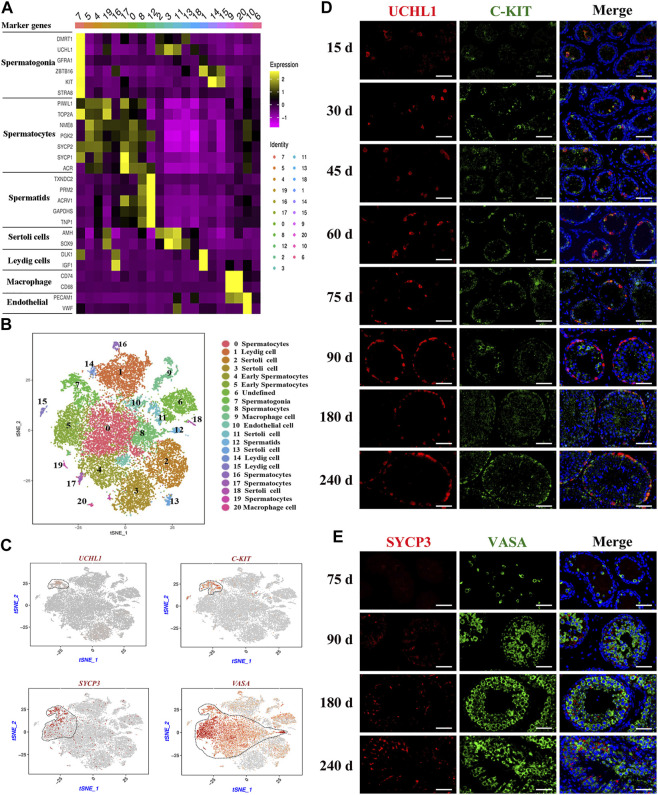
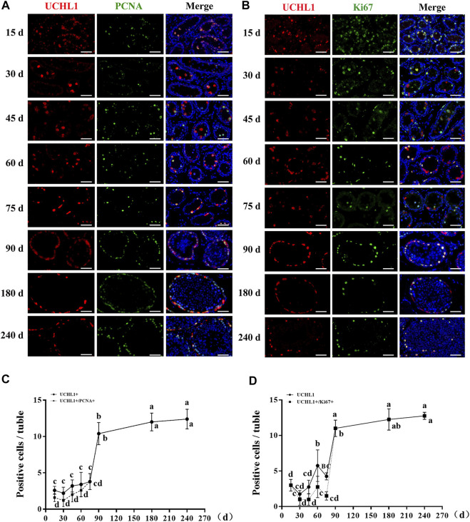
Spermatogenesis holds considerable promise for human-assisted reproduction and livestock breeding based on stem cells. It occurs in seminiferous tubules within the testis, which mainly comprise male germ cells and Sertoli cells. While the developmental progression of male germ cells and Sertoli cells has been widely reported in mice, much less is known in other large animal species, including dairy goats. In this study, we present the data of single cell RNA sequencing (scRNA-seq) for 25,373 cells from 45 (pre-puberty), 90 (puberty), and 180-day-old (post-puberty) dairy goat testes. We aimed to identify genes that are associated with key developmental events in male germ cells and Sertoli cells. We examined the development of spermatogenic cells and seminiferous tubules from 15, 30, 45, 60, 75, 90, 180, and 240-day-old buck goat testes. scRNA-seq clustering analysis of testicular cells from pre-puberty, puberty, and post-puberty goat testes revealed several cell types, including cell populations with characteristics of spermatogonia, early spermatocytes, spermatocytes, spermatids, Sertoli cells, Leydig cells, macrophages, and endothelial cells. We mapped the timeline for male germ cells development from spermatogonia to spermatids and identified gene signatures that define spermatogenic cell populations, such as AMH, SOHLH1, INHA, and ACTA2. Importantly, using immunofluorescence staining for different marker proteins (UCHL1, C-KIT, VASA, SOX9, AMH, and PCNA), we explored the proliferative activity and development of male germ cells and Sertoli cells. Moreover, we identified the expression patterns of potential key genes associated with the niche-related key pathways in male germ cells of dairy goats, including testosterone, retinoic acid, PDGF, FGF, and WNT pathways. In summary, our study systematically investigated the elaborate male germ cells and Sertoli cells developmental patterns in dairy goats that have so far remained largely unknown. This information represents a valuable resource for the establishment of goat male reproductive stem cells lines, induction of germ cell differentiation , and the exploration of sequential cell fate transition for spermatogenesis and testicular development at single-cell resolution.
基于干细胞的精子发生在人类辅助生殖和家畜育种方面具有巨大潜力。它发生在睾丸内的生精小管中,生精小管主要由雄性生殖细胞和支持细胞组成。虽然在小鼠中已广泛报道了雄性生殖细胞和支持细胞的发育进程,但在包括奶山羊在内的其他大型动物物种中,人们了解得却少得多。在本研究中,我们展示了来自45日龄(青春期前)、90日龄(青春期)和180日龄(青春期后)奶山羊睾丸的25373个细胞的单细胞RNA测序(scRNA-seq)数据。我们旨在鉴定与雄性生殖细胞和支持细胞关键发育事件相关的基因。我们研究了15、30、45、60、75、90、180和240日龄公山羊睾丸中生精细胞和生精小管的发育情况。对青春期前、青春期和青春期后奶山羊睾丸细胞进行的scRNA-seq聚类分析揭示了几种细胞类型,包括具有精原细胞、早期精母细胞、精母细胞、精子细胞、支持细胞、间质细胞、巨噬细胞和内皮细胞特征的细胞群体。我们绘制了从精原细胞到精子细胞的雄性生殖细胞发育时间线,并鉴定了定义生精细胞群体的基因特征,如抗苗勒管激素(AMH)、精子发生和睾丸生长相关蛋白1(SOHLH1)、抑制素A(INHA)和平滑肌肌动蛋白2(ACTA2)。重要的是,通过对不同标记蛋白(泛素羧基末端水解酶L1(UCHL1)、原癌基因c-KIT、血管生成素(VASA)、性别决定基因9(SOX9)、抗苗勒管激素(AMH)和增殖细胞核抗原(PCNA))进行免疫荧光染色,我们探究了雄性生殖细胞和支持细胞的增殖活性及发育情况。此外,我们确定了与奶山羊雄性生殖细胞中与微环境相关关键途径相关的潜在关键基因的表达模式,包括睾酮、视黄酸、血小板衍生生长因子(PDGF)、成纤维细胞生长因子(FGF)和WNT途径。总之,我们的研究系统地研究了奶山羊中迄今为止仍基本未知的精细雄性生殖细胞和支持细胞发育模式。这些信息是建立山羊雄性生殖干细胞系、诱导生殖细胞分化以及在单细胞分辨率下探索精子发生和睾丸发育的连续细胞命运转变的宝贵资源。