Kataoka Tomoya, Fukamoto Ayako, Hotta Yuji, Sanagawa Akimasa, Maeda Yasuhiro, Furukawa-Hibi Yoko, Kimura Kazunori
Department of Clinical Pharmaceutics, Graduate School of Medical Sciences, Nagoya City University, Nagoya, Japan; Department of Pharmacology, Kataoka's lab, Graduate School of Pharmaceutical Sciences, Chiba Institute of Science, Chiba, Japan.
Department of Hospital Pharmacy, Graduate School of Pharmaceutical Sciences, Nagoya City University, Nagoya, Japan.
Sex Med. 2022 Oct;10(5):100550. doi: 10.1016/j.esxm.2022.100550. Epub 2022 Aug 5.
Testosterone is an important hormone for the physical and mental health of men; however testosterone administration has also been suggested to adversely affect the cardiovascular system.
To investigate the effects of excessive testosterone administration on vascular endothelial and erectile function in rats.
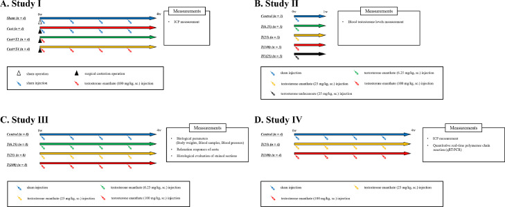
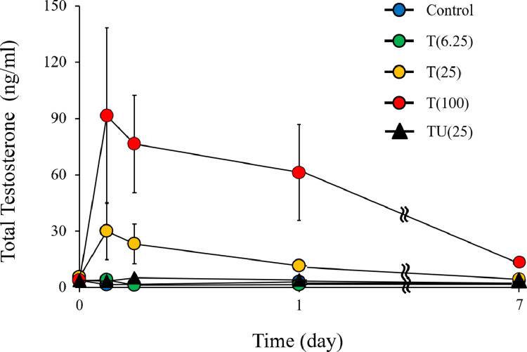
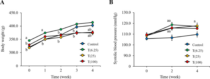
A total of seventy-five 12-week-old rats were divided into the following groups: Sham, castrated (Cast), castrated with subcutaneous administration of 100 mg/kg/month testosterone (Cast + T1), and castrated with subcutaneous administration of 100 mg/kg/week testosterone (Cast + T4). To observe the changes in testosterone level after the administration, rats were further divided into the following groups: control; T(6.25), wherein the rats were subcutaneously injected with 6.25 mg/kg testosterone; T(25) per week, wherein the rats were subcutaneously injected with 25 mg/kg testosterone per week; and T(100), wherein the rats were subcutaneously injected with 100 mg/kg testosterone per week. The relaxation responses of aorta were measured in these rats using standardized methods, and their erectile function was also evaluated. Statistical analysis of the obtained data was performed using two-way analysis of variance (ANOVA), Tukey-Kramer's multiple comparison test, or Student's t-test.
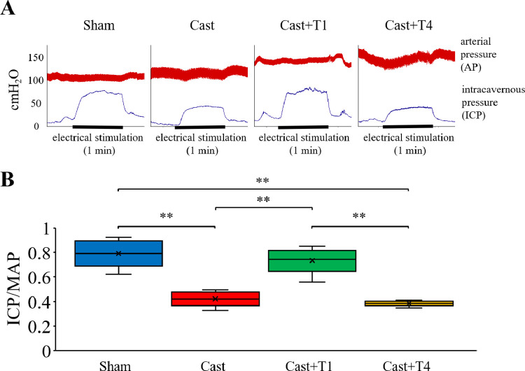
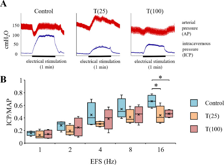
At the end of the study period, endothelial function was evaluated through measurement of isometric tension, while erectile function was assessed using intracavernosal pressure (ICP), mean arterial pressure (MAP), and the expression of endothelial nitric oxide synthase (eNOS), inducible nitric oxide synthase (iNOS), sirtuin 1 (Sirt1) and vascular endothelial growth factor A.
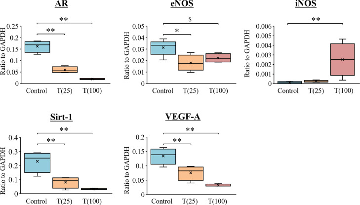
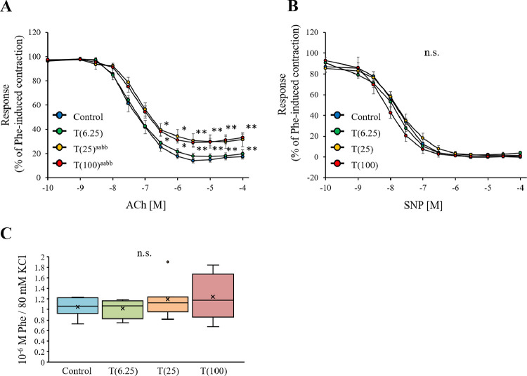
The ICP/MAP ratio in the Cast group (0.42 ± 0.04) was significantly lower than that in the Sham group (0.79 ± 0.07). The ICP/MAP ratio in the Cast + T1 group (0.73 ± 0.06) was significantly higher than that in the Cast group (P < .01) and that of the Cast + T4 (0.38 ± 0.01) group was unchanged (P > .05). The T(25) and T(100) groups exhibited significantly lower responses to ACh than the control group at 4 weeks (P < .01). Meanwhile, the ICP/MAP ratios in the T(25) group (0.44 ± 0.07) and T(100) group (0.47 ± 0.03) were significantly lower than that in the control group (0.67 ± 0.05) at stimulation frequencies of 16 Hz (P < .05). The expression of androgen receptor, Sirt1, and eNOS were significantly lower while that of iNOS was higher in the T(25) group compared with the control group (P < .05).
The results based on this animal model indicate that extremely high testosterone levels may affect endothelial and erectile function.
We found that high-dose testosterone administration decreased endothelial function in aorta and erectile function in rats. A major limitation of this study is that the blood concentration may not be representative of that in humans, and further research is needed.
The findings suggest that high doses of testosterone may cause endothelial dysfunction in the aorta and erectile dysfunction in rats and that the blood concentration should be monitored after testosterone administration. Kataoka T, Fukamoto A, Hotta Y, et al. Effect of High Testosterone Levels on Endothelial Function in Aorta and Erectile Function in Rats. Sex Med 2022;10:100550.
睾酮是对男性身心健康至关重要的一种激素;然而,也有人认为给予睾酮会对心血管系统产生不利影响。
研究过量给予睾酮对大鼠血管内皮功能和勃起功能的影响。
将75只12周龄的大鼠分为以下几组:假手术组(Sham)、去势组(Cast)、皮下注射100 mg/kg/月睾酮的去势组(Cast + T1)和皮下注射100 mg/kg/周睾酮的去势组(Cast + T4)。为观察给药后睾酮水平的变化,将大鼠进一步分为以下几组:对照组;T(6.25)组,即大鼠皮下注射6.25 mg/kg睾酮;T(25)每周组,即大鼠每周皮下注射25 mg/kg睾酮;以及T(100)组,即大鼠每周皮下注射100 mg/kg睾酮。采用标准化方法测量这些大鼠主动脉的舒张反应,并评估其勃起功能。使用双向方差分析(ANOVA)、Tukey-Kramer多重比较检验或Student t检验对所得数据进行统计分析。
研究期末,通过测量等长张力评估内皮功能,同时使用海绵体内压(ICP)、平均动脉压(MAP)以及内皮型一氧化氮合酶(eNOS)、诱导型一氧化氮合酶(iNOS)、沉默调节蛋白1(Sirt1)和血管内皮生长因子A的表达评估勃起功能。
去势组(Cast)的ICP/MAP比值(0.42±0.04)显著低于假手术组(Sham)(0.79±0.07)。Cast + T1组的ICP/MAP比值(0.73±0.06)显著高于去势组(P <.01),而Cast + T4组(0.38±0.01)的比值未改变(P>.05)。在4周时,T(25)组和T(100)组对乙酰胆碱(ACh)的反应显著低于对照组(P <.01)。同时,在16 Hz刺激频率下,T(25)组(0.44±0.07)和T(100)组(0.47±0.03)的ICP/MAP比值显著低于对照组(0.67±0.05)(P <.05)。与对照组相比,T(25)组雄激素受体、Sirt1和eNOS的表达显著降低,而iNOS的表达升高(P <.05)。
基于该动物模型的结果表明,极高的睾酮水平可能会影响内皮功能和勃起功能。
我们发现高剂量给予睾酮会降低大鼠主动脉的内皮功能和勃起功能。本研究的一个主要局限性是血液浓度可能不代表人类的情况,需要进一步研究。
研究结果表明,高剂量睾酮可能导致大鼠主动脉内皮功能障碍和勃起功能障碍,且在给予睾酮后应监测血液浓度。片冈T、深本A、堀田Y等。高睾酮水平对大鼠主动脉内皮功能和勃起功能的影响。性医学2022;10:100550。