Biomedical and Data Sciences Division, CFD Research Corporation, Huntsville, AL 35806, USA.
Department of Bioengineering, Temple University, Philadelphia, PA 19122, USA.
Int J Mol Sci. 2022 Jul 29;23(15):8399. doi: 10.3390/ijms23158399.
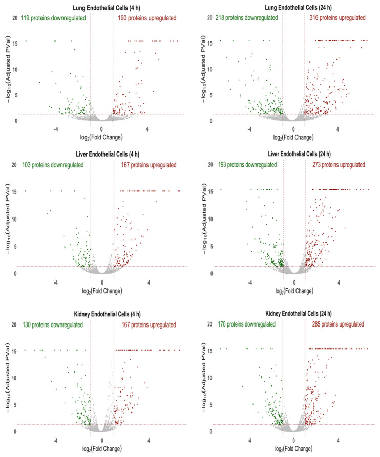
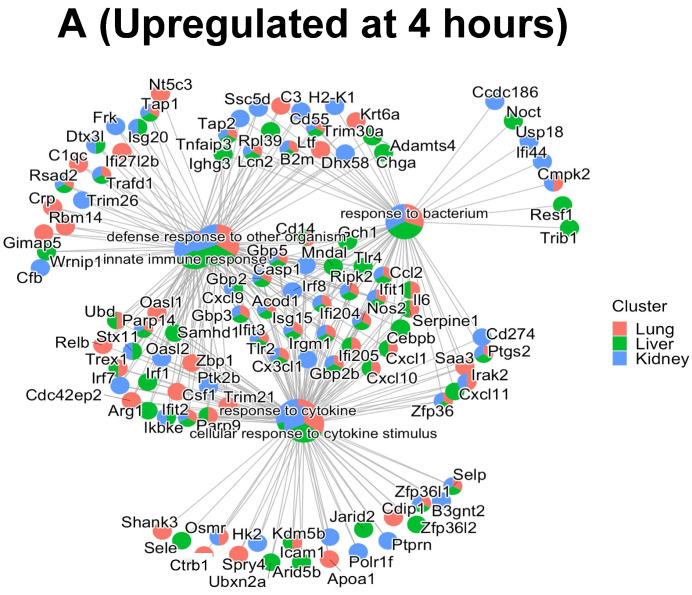
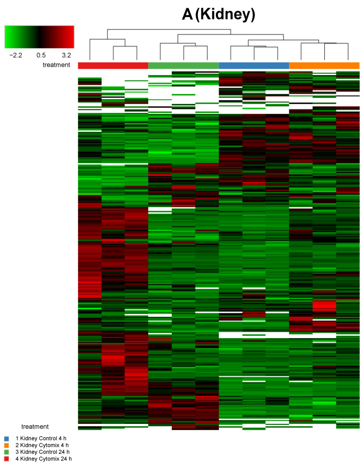
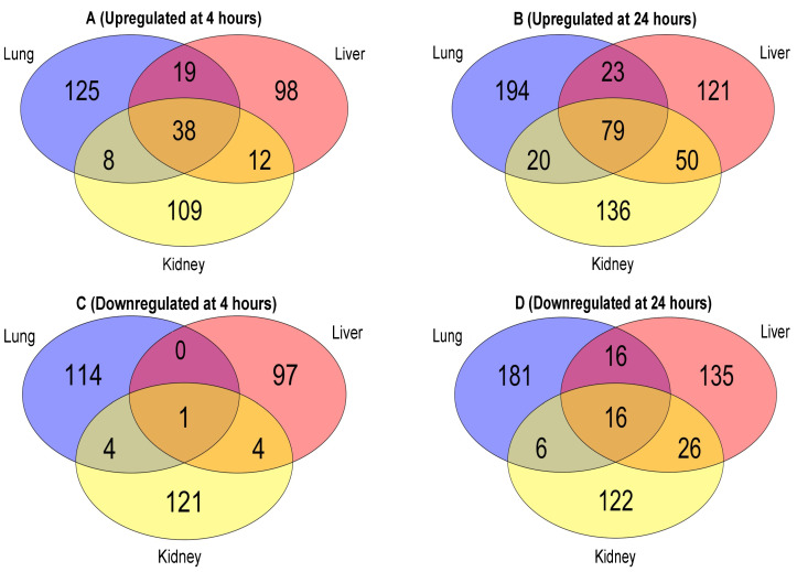
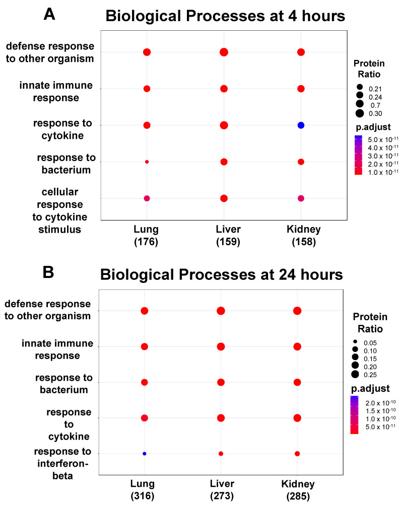
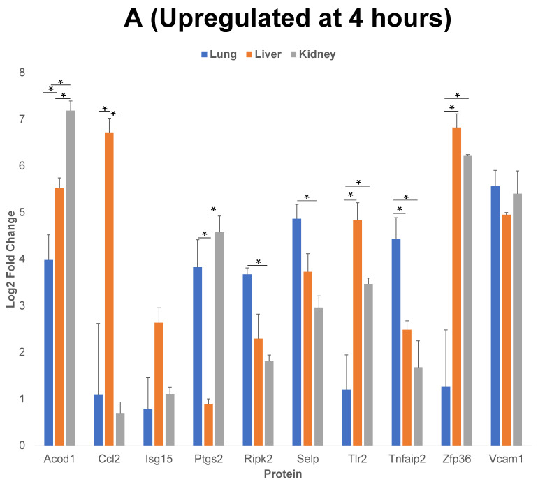
A key aspect of cytokine-induced changes as observed in sepsis is the dysregulated activation of endothelial cells (ECs), initiating a cascade of inflammatory signaling leading to leukocyte adhesion/migration and organ damage. The therapeutic targeting of ECs has been hampered by concerns regarding organ-specific EC heterogeneity and their response to inflammation. Using in vitro and in silico analysis, we present a comprehensive analysis of the proteomic changes in mouse lung, liver and kidney ECs following exposure to a clinically relevant cocktail of proinflammatory cytokines. Mouse lung, liver and kidney ECs were incubated with TNF-α/IL-1β/IFN-γ for 4 or 24 h to model the cytokine-induced changes. Quantitative label-free global proteomics and bioinformatic analysis performed on the ECs provide a molecular framework for the EC response to inflammatory stimuli over time and organ-specific differences. Gene Ontology and PANTHER analysis suggest why some organs are more susceptible to inflammation early on, and show that, as inflammation progresses, some protein expression patterns become more uniform while additional organ-specific proteins are expressed. These findings provide an in-depth understanding of the molecular changes involved in the EC response to inflammation and can support the development of drugs targeting ECs within different organs. Data are available via ProteomeXchange (identifier PXD031804).
细胞因子诱导的变化是脓毒症的一个关键方面,其特征是内皮细胞(ECs)的失调激活,引发一连串的炎症信号级联反应,导致白细胞黏附和迁移以及器官损伤。由于担心内皮细胞的器官特异性异质性及其对炎症的反应,针对内皮细胞的治疗靶向受到了阻碍。我们使用体外和计算分析,对小鼠肺、肝和肾 ECs 在暴露于临床相关促炎细胞因子混合物后的蛋白质组变化进行了全面分析。用 TNF-α/IL-1β/IFN-γ孵育小鼠肺、肝和肾 EC 4 或 24 小时,以模拟细胞因子诱导的变化。对 EC 进行定量无标记全局蛋白质组学和生物信息学分析,为 EC 对炎症刺激的时间和器官特异性差异的反应提供了分子框架。GO 和 PANTHER 分析表明为什么一些器官早期更容易受到炎症的影响,并表明随着炎症的进展,一些蛋白质表达模式变得更加一致,而其他器官特异性蛋白质则被表达。这些发现深入了解了 EC 对炎症反应所涉及的分子变化,并可为针对不同器官的 EC 开发药物提供支持。数据可通过 ProteomeXchange(标识符 PXD031804)获得。