Raschke F, Witzmann K, Seidlitz A, Wesemann T, Jentsch C, Platzek I, van den Hoff J, Kotzerke J, Beuthien-Baumann B, Baumann M, Linn J, Krause M, Troost E G C
Institute of Radiooncology - OncoRay, Helmholtz-Zentrum Dresden-Rossendorf, Rossendorf, Germany.
OncoRay - National Center for Radiation Research in Oncology, Faculty of Medicine and University Hospital Carl Gustav Carus, Technische Universität Dresden, Helmholtz-Zentrum Dresden - Rossendorf, Dresden, Germany.
Clin Transl Radiat Oncol. 2022 Jul 20;36:99-105. doi: 10.1016/j.ctro.2022.07.003. eCollection 2022 Sep.
Radiotherapy (RT) is an adjuvant treatment option for glioma patients. Side effects include tissue atrophy, which might be a contributing factor to neurocognitive decline after treatment. The goal of this study was to determine potential atrophy of the hippocampus, amygdala, thalamus, putamen, pallidum and caudate nucleus in glioma patients having undergone magnetic resonance imaging (MRI) before and after RT.
Subcortical volumes were measured using T1-weighted MRI from patients before RT (N = 91) and from longitudinal follow-ups acquired in three-monthly intervals (N = 349). The volumes were normalized to the baseline values, while excluding structures touching the clinical target volume (CTV) or abnormal tissue seen on FLAIR imaging. A multivariate linear effects model was used to determine if time after RT and mean RT dose delivered to the corresponding structures were significant predictors of tissue atrophy.
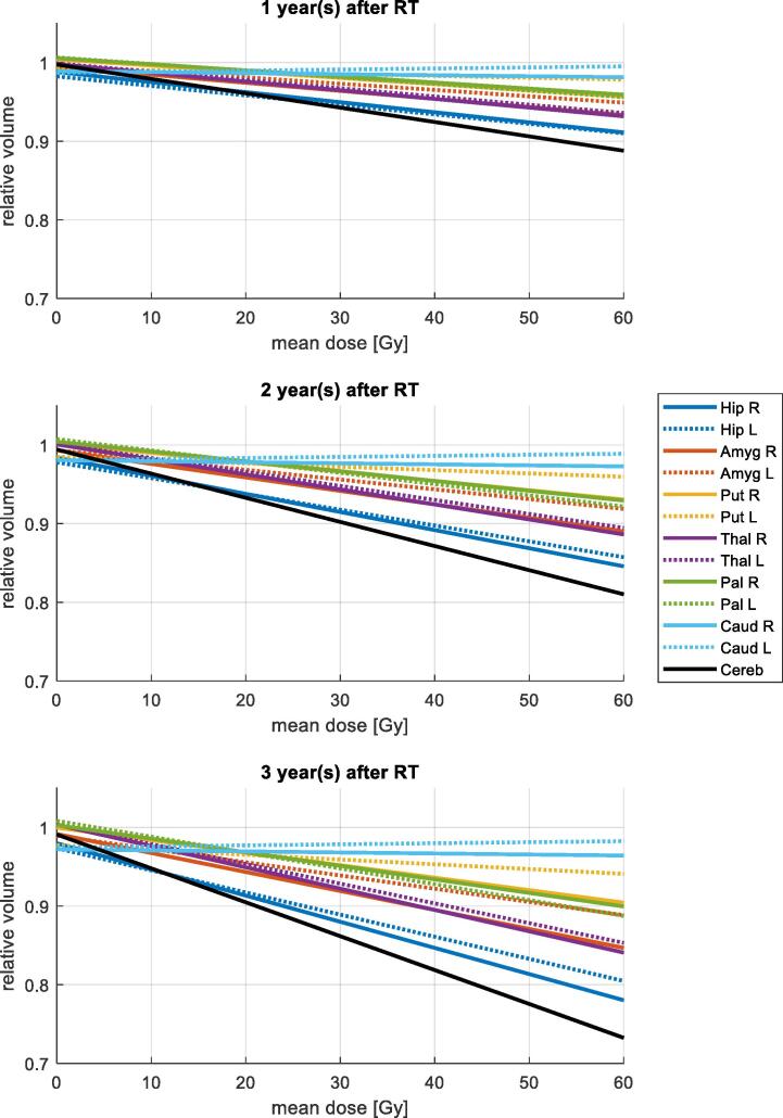
The hippocampus, amygdala, thalamus, putamen, and pallidum showed significant atrophy after RT as function of both time after RT and mean RT dose delivered to the corresponding structure. Only the caudate showed no dose or time dependant atrophy. Conversely, the hippocampus was the structure with the highest atrophy rate of 5.2 % after one year and assuming a mean dose of 30 Gy.
The hippocampus showed the highest atrophy rates followed by the thalamus and the amygdala. The subcortical structures here found to decrease in volume indicative of radiosensitivity should be the focus of future studies investigating the relationship between neurocognitive decline and RT.
放射治疗(RT)是胶质瘤患者的一种辅助治疗选择。其副作用包括组织萎缩,这可能是治疗后神经认知功能下降的一个促成因素。本研究的目的是确定接受放射治疗(RT)前后进行磁共振成像(MRI)检查的胶质瘤患者海马体、杏仁核、丘脑、壳核、苍白球和尾状核的潜在萎缩情况。
使用T1加权MRI测量RT前患者(N = 91)以及每三个月进行一次纵向随访患者(N = 349)的皮质下体积。将体积标准化为基线值,同时排除接触临床靶体积(CTV)的结构或液体衰减反转恢复序列(FLAIR)成像上可见的异常组织。使用多元线性效应模型来确定RT后时间以及输送到相应结构的平均RT剂量是否是组织萎缩的显著预测因素。
RT后,海马体、杏仁核、丘脑、壳核和苍白球均出现显著萎缩,这是RT后时间和输送到相应结构的平均RT剂量的函数。只有尾状核未表现出剂量或时间依赖性萎缩。相反,海马体是萎缩率最高的结构,在一年后萎缩率为5.2%,假设平均剂量为30 Gy。
海马体萎缩率最高,其次是丘脑和杏仁核。这里发现体积减小表明具有放射敏感性的皮质下结构应成为未来研究神经认知功能下降与RT之间关系的重点。