Bishop Courtney A, Newbould Rexford D, Lee Jean Sz, Honeyfield Lesley, Quest Rebecca, Colasanti Alessandro, Ali Rehiana, Mattoscio Miriam, Cortese Antonio, Nicholas Richard, Matthews Paul M, Muraro Paolo A, Waldman Adam D
Imanova Centre for Imaging Sciences, London, UK.
Division of Brain Sciences, Imperial College London, UK; Department of Imaging, Imperial College Healthcare NHS Trust, London, UK.
Neuroimage Clin. 2016 Nov 9;13:9-15. doi: 10.1016/j.nicl.2016.11.005. eCollection 2017.
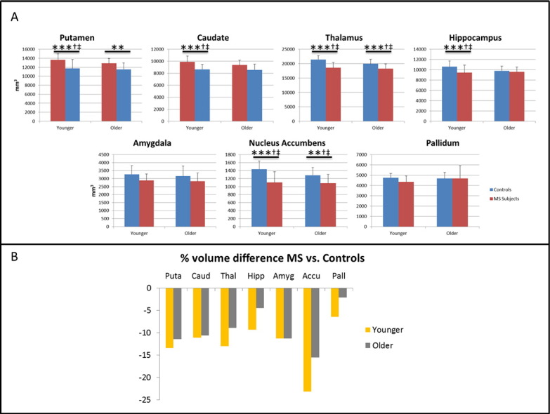
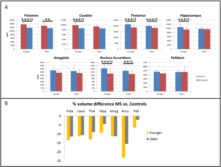
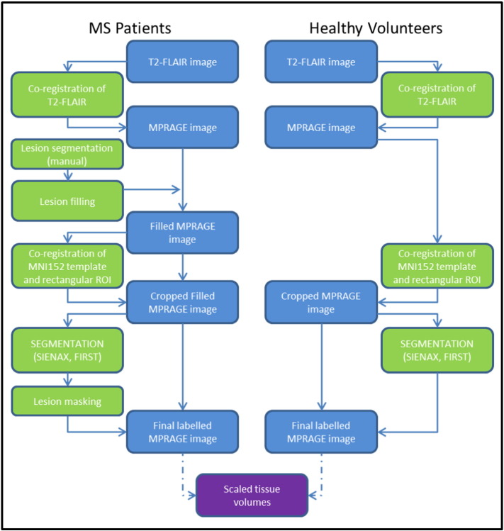
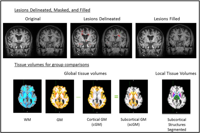
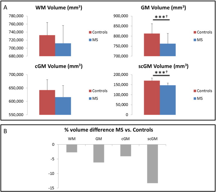
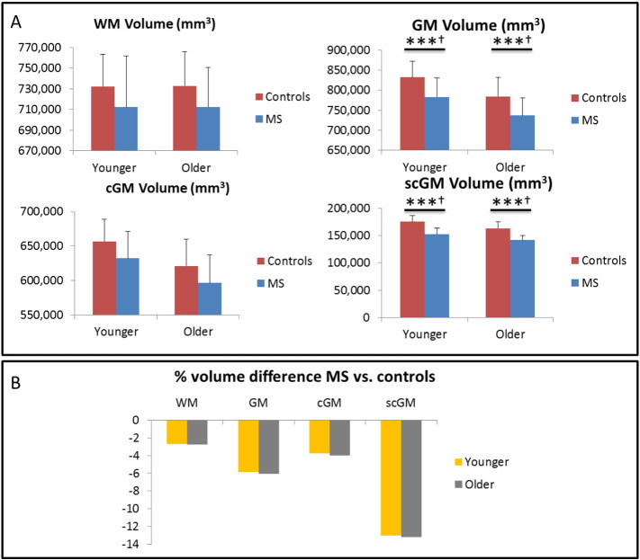
Age of onset in multiple sclerosis (MS) exerts an influence on the course of disease. This study examined whether global and regional brain volumes differed between "younger" and "older" onset MS subjects who were matched for short disease duration, mean 1.9 years and burden as measured by the MS Severity Score and relapses. 21 younger-onset MS subjects (age 30.4 ± 3.2 years) were compared with 17 older-onset (age 48.7 ± 3.3 years) as well as age-matched controls ( = 31, 31.9 ± 3.5 years and = 21, 47.3 ± 4.0 years). All subjects underwent 3D volumetric T1 and T2-FLAIR imaging. White matter (WM) and grey matter (GM) lesions were outlined manually. Lesions were filled prior to tissue and structural segmentation to reduce classification errors. Volume loss versus control was predominantly in the subcortical GM, at > 13% loss. Younger and older-onset MS subjects had similar, strong excess loss in the putamen, thalamus, and nucleus accumbens. No excess loss was detected in the amygdala or pallidum. The hippocampus and caudate showed significant excess loss in the younger group ( < 0.001) and a strong trend in the older-onset group. These results provide a potential imaging correlate of published neuropsychological studies that reported the association of younger age at disease onset with impaired cognitive performance, including decreased working memory.
多发性硬化症(MS)的发病年龄会对疾病进程产生影响。本研究调查了疾病持续时间短(平均1.9年)且经MS严重程度评分和复发次数衡量负担相当的“早发型”和“晚发型”MS患者在全脑和局部脑容量上是否存在差异。将21例早发型MS患者(年龄30.4±3.2岁)与17例晚发型患者(年龄48.7±3.3岁)以及年龄匹配的对照组(n = 31,31.9±3.5岁和n = 21,47.3±4.0岁)进行比较。所有受试者均接受了三维容积T1和T2-FLAIR成像。对白质(WM)和灰质(GM)病变进行手动勾勒。在进行组织和结构分割之前填充病变,以减少分类错误。与对照组相比,体积损失主要发生在皮质下GM,损失超过13%。早发型和晚发型MS患者在壳核、丘脑和伏隔核中均有类似的、明显的过度损失。杏仁核或苍白球未检测到过度损失。海马体和尾状核在年轻组中显示出显著的过度损失(p<0.001),在晚发型组中有强烈趋势。这些结果为已发表的神经心理学研究提供了潜在的影像学关联,这些研究报告了疾病发病年龄较小与认知功能受损(包括工作记忆下降)之间的关联。