Nagtegaal Steven H J, David Szabolcs, Philippens Marielle E P, Snijders Tom J, Leemans Alexander, Verhoeff Joost J C
Department of Radiation Oncology, University Medical Center Utrecht, HP Q 00.3.11, PO Box 85500, 3508 GA Utrecht, the Netherlands.
UMC Utrecht Brain Center, Department of Neurology & Neurosurgery, University Medical Center Utrecht, HP L 01.310, PO Box 85500, 3508 GA Utrecht, the Netherlands.
Clin Transl Radiat Oncol. 2020 Nov 15;26:35-41. doi: 10.1016/j.ctro.2020.11.005. eCollection 2021 Jan.
The relation between radiotherapy (RT) dose to the brain and morphological changes in healthy tissue has seen recent increased interest. There already is evidence for changes in the cerebral cortex and white matter, as well as selected subcortical grey matter (GM) structures. We studied this relation in all deep GM structures, to help understand the aetiology of post-RT neurocognitive symptoms.
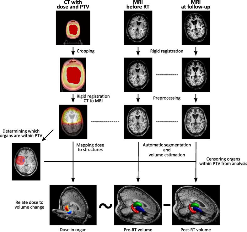
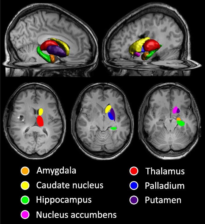
We selected 31 patients treated with RT for grade II-IV glioma. Pre-RT and 1 year post-RT 3D T1-weighted MRIs were automatically segmented, and the changes in volume of the following structures were assessed: amygdala, nucleus accumbens, caudate nucleus, hippocampus, globus pallidus, putamen, and thalamus. The volumetric changes were related to the mean RT dose received by each structure. Hippocampal volumes were entered into a population-based nomogram to estimate hippocampal age.
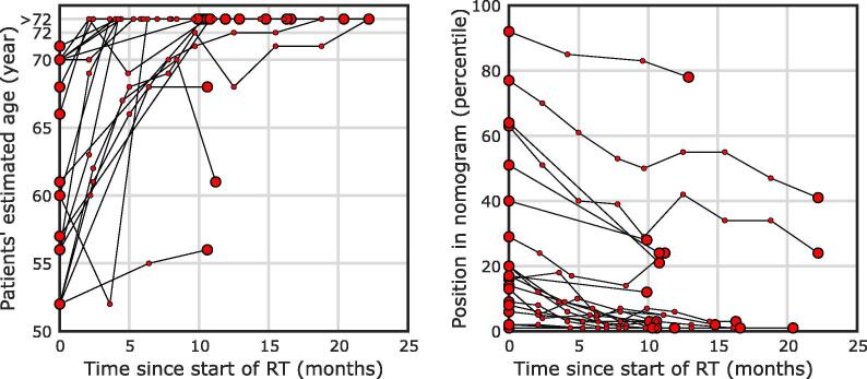
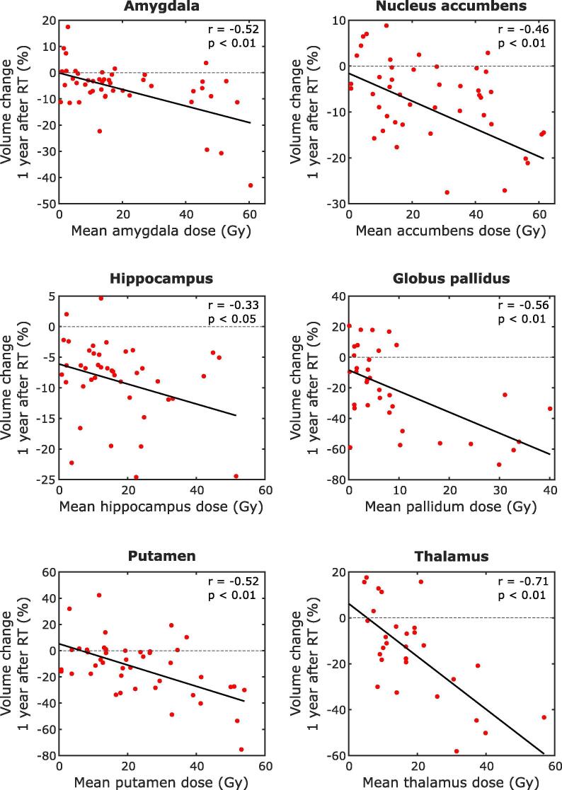
A significant relation between RT dose and volume loss was seen in all examined structures, except the caudate nucleus. The volume loss rates ranged from 0.16 to 1.37%/Gy, corresponding to 4.9-41.2% per 30 Gy. Hippocampal age, as derived from the nomogram, was seen to increase by a median of 11 years.
Almost all subcortical GM structures are susceptible to radiation-induced volume loss, with higher volume loss being observed with increasing dose. Volume loss of these structures is associated with neurological deterioration, including cognitive decline, in neurodegenerative diseases. To support a causal relationship between radiation-induced deep GM loss and neurocognitive functioning in glioma patients, future studies are needed that directly correlate volumetrics to clinical outcomes.
脑部放疗(RT)剂量与健康组织形态学变化之间的关系近来受到越来越多的关注。已有证据表明大脑皮层、白质以及部分皮质下灰质(GM)结构会发生变化。我们对所有深部GM结构中的这种关系进行了研究,以帮助理解放疗后神经认知症状的病因。
我们选取了31例接受RT治疗的II-IV级胶质瘤患者。对放疗前及放疗后1年的三维T1加权磁共振成像进行自动分割,并评估以下结构的体积变化:杏仁核、伏隔核、尾状核、海马体、苍白球、壳核和丘脑。体积变化与每个结构所接受的平均RT剂量相关。将海马体体积输入基于人群的列线图以估计海马体年龄。
除尾状核外,在所有检查的结构中均观察到RT剂量与体积损失之间存在显著关系。体积损失率范围为0.16%至1.37%/Gy,相当于每30 Gy损失4.9%至41.2%。从列线图得出的海马体年龄中位数增加了11岁。
几乎所有皮质下GM结构都易受辐射诱导的体积损失影响,且剂量增加时体积损失更大。这些结构的体积损失与神经退行性疾病中的神经功能恶化有关,包括认知衰退。为了支持辐射诱导的深部GM损失与胶质瘤患者神经认知功能之间的因果关系,未来需要进行将体积测量与临床结果直接关联的研究。