Mohajeri Mahsa, Khazaei Sara, Vafaee Fariborz, Firouz Farnaz, Ghorbani Gholiabad Somayeh, Shisheian Arash
Department of Prosthodontics, School of Dentistry, Tehran University of Medical Sciences, Tehran, Iran.
Department of Prosthodontics, Dental Faculty, Hamadan University of Medical Sciences, Hamadan, Iran.
Front Dent. 2021 Sep 5;18:31. doi: 10.18502/fid.v18i31.7236. eCollection 2021.
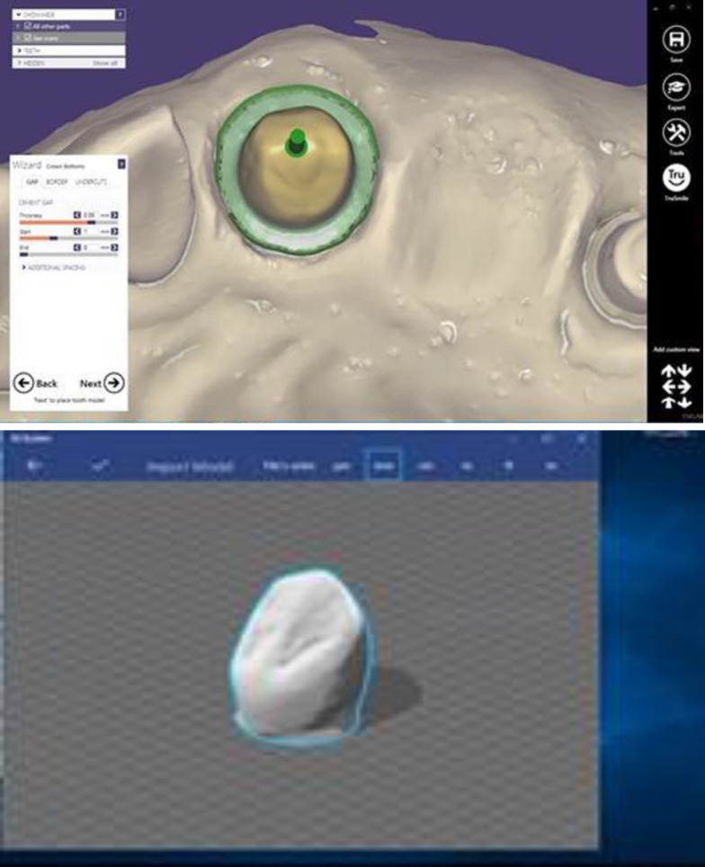
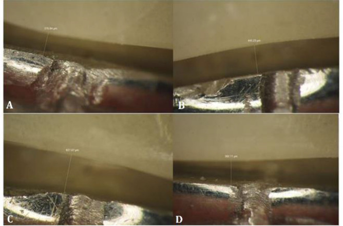
This study aimed to compare the marginal fit of temporary restorations fabricated by the conventional chairside method, 3D printing, and milling. In this in vitro, experimental study, 14 temporary restorations were conventionally fabricated over an implant abutment and analog that had been mounted in a phantom model at the site of canine tooth, using auto-polymerizing acrylic resin and putty index. In digital manufacturing, the original model was scanned, and the final restoration was designed. Fourteen temporary restorations were milled out of polymethyl methacrylate (PMMA) blocks, and 14 others were printed by a 3D printer. Temporary crowns were placed on the abutment, and images were obtained from specific areas under a stereomicroscope at x100 magnification to measure the amount of marginal gap. Data were analyzed using one-way ANOVA and Tukey's test (α=0.05). The mean marginal gap values for the temporary crowns in the 3D printing, milling, and chairside groups were 91.40, 75.28 and 51.23 µm, respectively. The crowns that were conventionally fabricated chairside exhibited the lowest marginal gap, and the difference in this respect was significant among the three groups (P<0.05). Temporary crowns fabricated by the chairside method showed significantly smaller marginal gap; however, the marginal gap of all three groups was within the clinically acceptable range.
本研究旨在比较采用传统椅旁法、3D打印和铣削制作的临时修复体的边缘适合性。在这项体外实验研究中,在犬齿部位安装于模型中的种植体基台及代型上,使用自凝丙烯酸树脂和印模膏,通过传统方法制作14个临时修复体。在数字制造中,扫描原始模型并设计最终修复体。用聚甲基丙烯酸甲酯(PMMA)块铣削出14个临时修复体,另外14个由3D打印机打印。将临时冠放置在基台上,在体视显微镜下以100倍放大倍数从特定区域获取图像,以测量边缘间隙量。使用单因素方差分析和Tukey检验(α = 0.05)分析数据。3D打印组、铣削组和椅旁组临时冠的平均边缘间隙值分别为91.40、75.28和51.23µm。传统椅旁制作的冠边缘间隙最小,三组在这方面的差异具有统计学意义(P < 0.05)。椅旁法制作的临时冠边缘间隙明显更小;然而,所有三组的边缘间隙均在临床可接受范围内。