Lin Bingbing, Zhang Lanlan, Yin Xiaolong, Chen Xiaocheng, Ruan Chendong, Wu Tiecheng, Liu Zhizhen, Huang Jia
College of Rehabilitation Medicine, Fujian University of Traditional Chinese Medicine, Fuzhou, China.
TCM Rehabilitation Research Center of State Administration of Traditional Chinese Medicine (SATCM), Fujian University of Traditional Chinese Medicine, Fuzhou, China.
Front Neurosci. 2022 Jul 29;16:968767. doi: 10.3389/fnins.2022.968767. eCollection 2022.
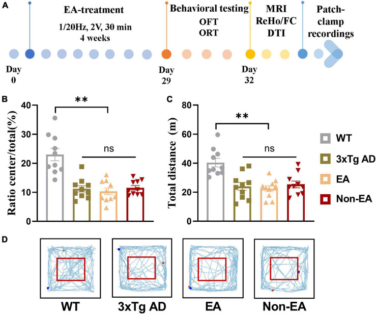
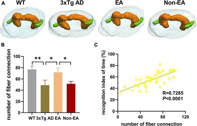
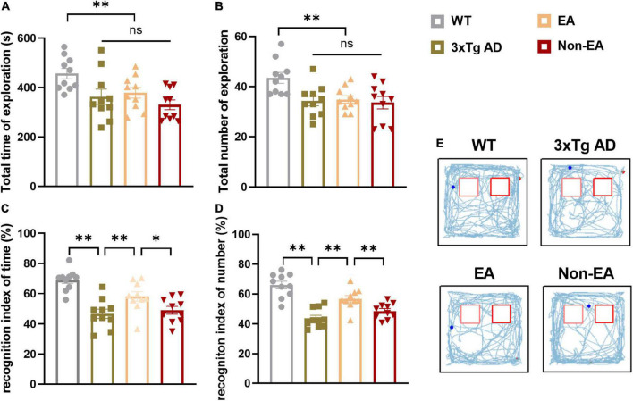
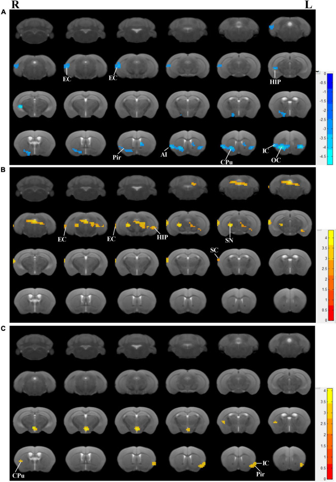
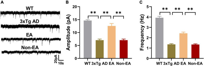
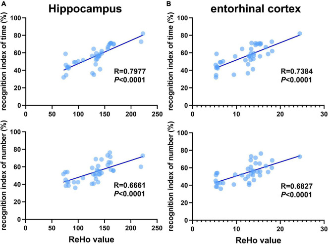
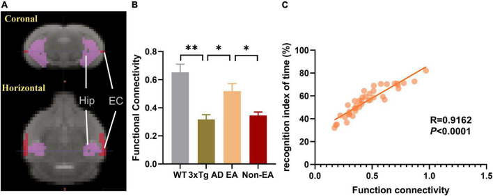
Memory loss and aberrant neuronal network activity are part of the earliest hallmarks of Alzheimer's disease (AD). Electroacupuncture (EA) has been recognized as a cognitive stimulation for its effects on memory disorder, but whether different brain regions or neural circuits contribute to memory recovery in AD remains unknown. Here, we found that memory deficit was ameliorated in 3×Tg-AD mice with EA-treatment, as shown by the increased number of exploring and time spent in the novel object. In addition, reduced locomotor activity was observed in 3×Tg-AD mice, but no significant alteration was seen in the EA-treated mice. Based on the functional magnetic resonance imaging, the regional spontaneous activity alterations of 3×Tg-AD were mainly concentrated in the accumbens nucleus, auditory cortex, caudate putamen, entorhinal cortex (EC), hippocampus, insular cortex, subiculum, temporal cortex, visual cortex, and so on. While EA-treatment prevented the chaos of brain activity in parts of the above regions, such as the auditory cortex, EC, hippocampus, subiculum, and temporal cortex. And then we used the whole-cell voltage-clamp recording to reveal the neurotransmission in the hippocampus, and found that EA-treatment reversed the synaptic spontaneous release. Since the hippocampus receives most of the projections of the EC, the hippocampus-EC circuit is one of the neural circuits related to memory impairment. We further applied diffusion tensor imaging (DTI) tracking and functional connectivity, and found that hypo-connected between the hippocampus and EC with EA-treatment. These data indicate that the hippocampus-EC connectivity is responsible for the recognition memory deficit in the AD mice with EA-treatment, and provide novel insight into potential therapies for memory loss in AD.
记忆丧失和异常的神经网络活动是阿尔茨海默病(AD)最早的特征之一。电针(EA)因其对记忆障碍的影响而被认为是一种认知刺激,但不同脑区或神经回路是否有助于AD患者的记忆恢复仍不清楚。在这里,我们发现经EA治疗的3×Tg-AD小鼠的记忆缺陷得到改善,如在新物体中探索的次数增加和停留时间延长所示。此外,在3×Tg-AD小鼠中观察到运动活动减少,但在经EA治疗的小鼠中未观察到明显变化。基于功能磁共振成像,3×Tg-AD小鼠的区域自发活动改变主要集中在伏隔核、听觉皮层、尾状壳核、内嗅皮层(EC)、海马体、岛叶皮层、下托、颞叶皮层、视觉皮层等。而EA治疗可防止上述部分区域(如听觉皮层、EC、海马体、下托和颞叶皮层)的脑活动紊乱。然后我们使用全细胞膜片钳记录来揭示海马体中的神经传递,发现EA治疗可逆转突触自发释放。由于海马体接收EC的大部分投射,海马体-EC回路是与记忆障碍相关的神经回路之一。我们进一步应用扩散张量成像(DTI)追踪和功能连接性,发现经EA治疗后海马体与EC之间的连接减弱。这些数据表明,海马体-EC连接性是经EA治疗的AD小鼠识别记忆缺陷的原因,并为AD记忆丧失的潜在治疗提供了新的见解。