• 文献检索
  • 文档翻译
  • 深度研究
  • 学术资讯
  • Suppr Zotero 插件Zotero 插件
  • 邀请有礼
  • 套餐&价格
  • 历史记录
应用&插件
Suppr Zotero 插件Zotero 插件浏览器插件Mac 客户端Windows 客户端微信小程序
定价
高级版会员购买积分包购买API积分包
服务
文献检索文档翻译深度研究API 文档MCP 服务
关于我们
关于 Suppr公司介绍联系我们用户协议隐私条款
关注我们

Suppr 超能文献

核心技术专利:CN118964589B侵权必究
粤ICP备2023148730 号-1Suppr @ 2026

文献检索

告别复杂PubMed语法,用中文像聊天一样搜索,搜遍4000万医学文献。AI智能推荐,让科研检索更轻松。

立即免费搜索

文件翻译

保留排版,准确专业,支持PDF/Word/PPT等文件格式,支持 12+语言互译。

免费翻译文档

深度研究

AI帮你快速写综述,25分钟生成高质量综述,智能提取关键信息,辅助科研写作。

立即免费体验

MKK6 缺乏通过 MKK3-p38γ/δ-mTOR 的过度激活促进心脏功能障碍。

MKK6 deficiency promotes cardiac dysfunction through MKK3-p38γ/δ-mTOR hyperactivation.

机构信息

Centro Nacional de Investigaciones Cardiovasculares, Madrid, Spain.

CIBER de Enfermedades Cardiovasculares, Madrid, Spain.

出版信息

Elife. 2022 Aug 16;11:e75250. doi: 10.7554/eLife.75250.

DOI:10.7554/eLife.75250
PMID:35971771
原文链接:https://pmc.ncbi.nlm.nih.gov/articles/PMC9381040/
Abstract

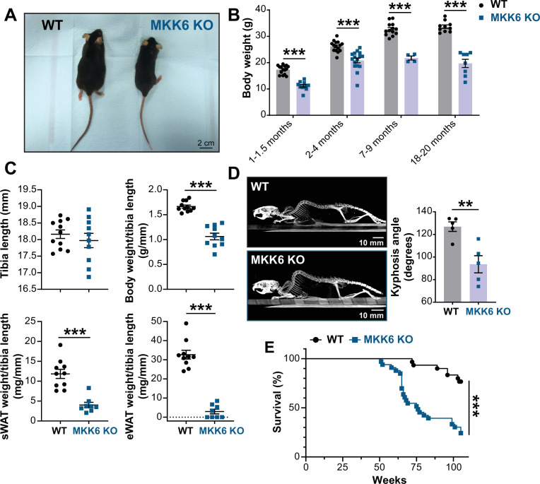
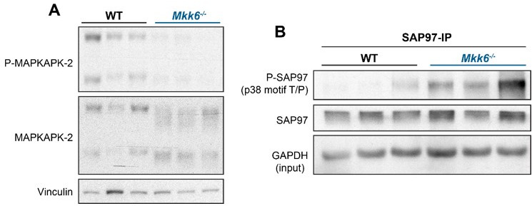
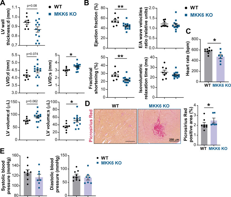
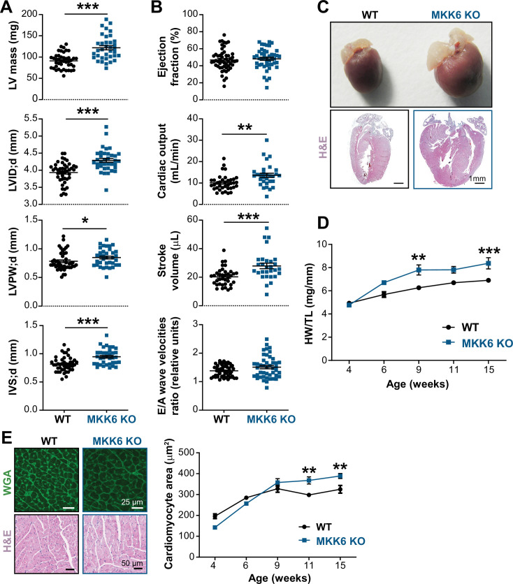
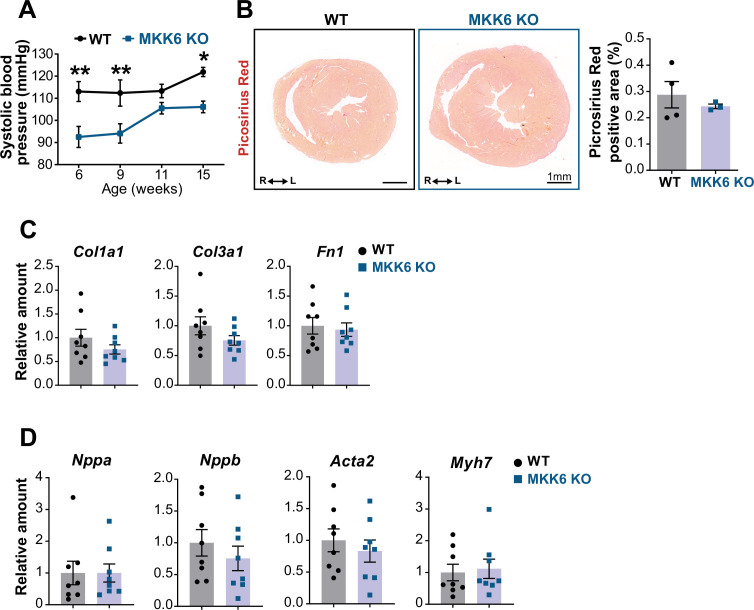
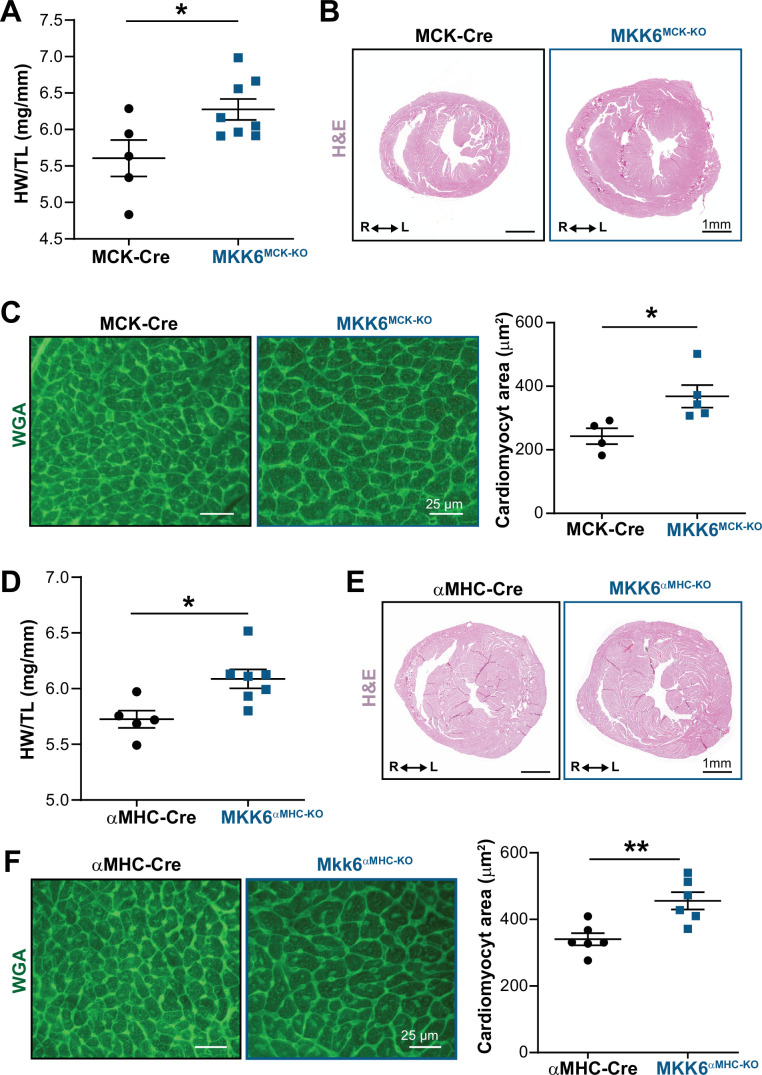
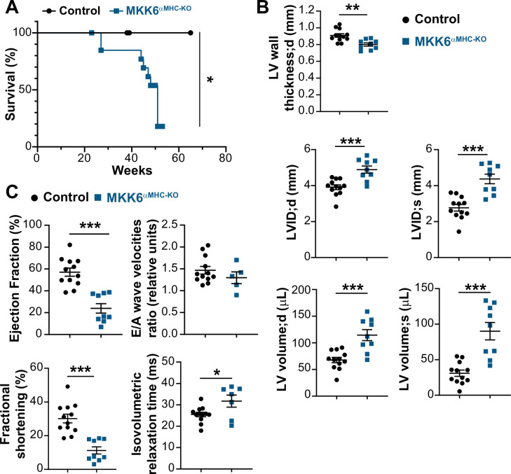
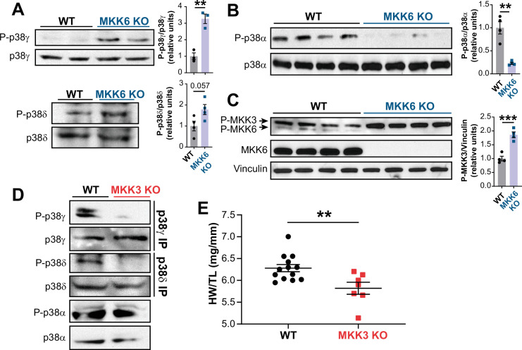
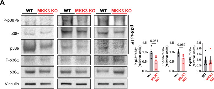
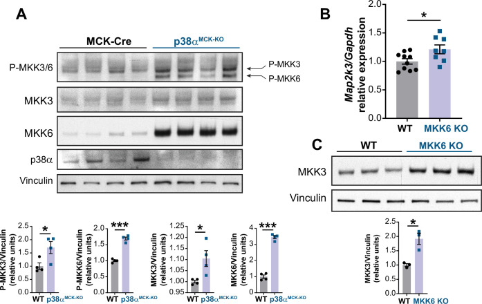
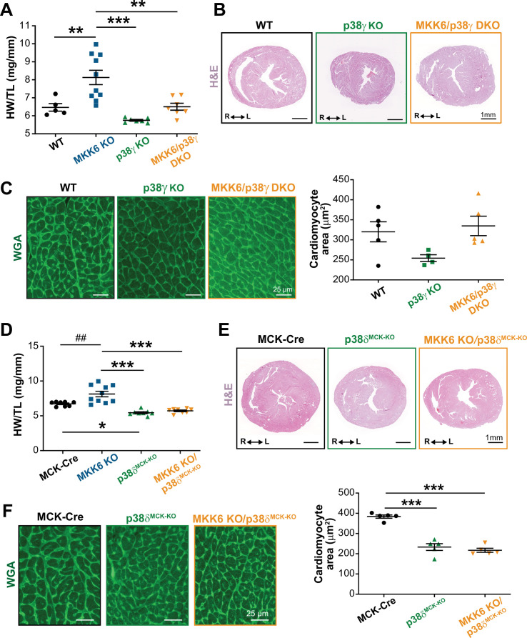
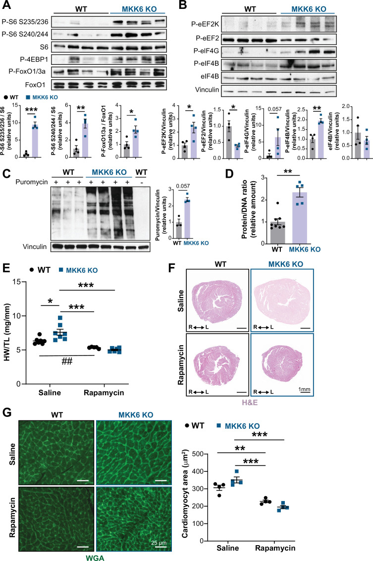
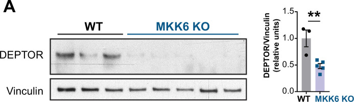
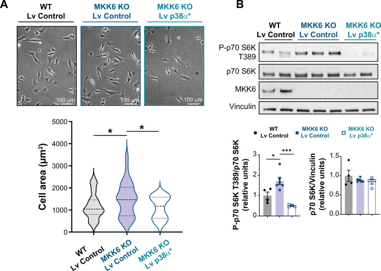
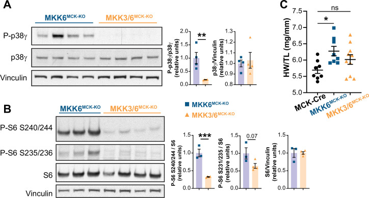
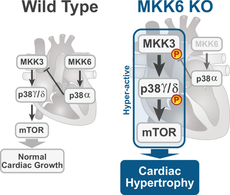
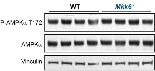
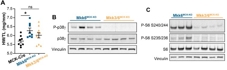
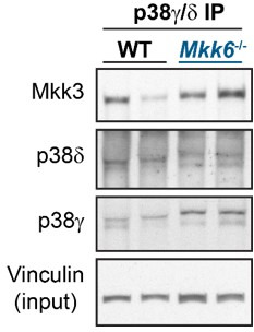
Stress-activated p38 kinases control a plethora of functions, and their dysregulation has been linked to the development of steatosis, obesity, immune disorders, and cancer. Therefore, they have been identified as potential targets for novel therapeutic strategies. There are four p38 family members (p38α, p38β, p38γ, and p38δ) that are activated by MKK3 and MKK6. Here, we demonstrate that lack of MKK6 reduces the lifespan in mice. Longitudinal study of cardiac function in MKK6 KO mice showed that young mice develop cardiac hypertrophy which progresses to cardiac dilatation and fibrosis with age. Mechanistically, lack of MKK6 blunts p38α activation while causing MKK3-p38γ/δ hyperphosphorylation and increased mammalian target of rapamycin (mTOR) signaling, resulting in cardiac hypertrophy. Cardiac hypertrophy in MKK6 KO mice is reverted by knocking out either p38γ or p38δ or by inhibiting the mTOR pathway with rapamycin. In conclusion, we have identified a key role for the MKK3/6-p38γ/δ pathway in the development of cardiac hypertrophy, which has important implications for the clinical use of p38α inhibitors in the long-term treatment since they might result in cardiotoxicity.

摘要

应激激活的 p38 激酶控制着大量的功能,其失调与脂肪变性、肥胖、免疫紊乱和癌症的发展有关。因此,它们被认为是新的治疗策略的潜在靶点。有四个 p38 家族成员(p38α、p38β、p38γ 和 p38δ)被 MKK3 和 MKK6 激活。在这里,我们证明缺乏 MKK6 会缩短小鼠的寿命。对 MKK6 KO 小鼠心脏功能的纵向研究表明,年轻小鼠会发生心脏肥大,随着年龄的增长,会进展为心脏扩张和纤维化。从机制上讲,缺乏 MKK6 会抑制 p38α 的激活,同时导致 MKK3-p38γ/δ 的过度磷酸化和哺乳动物雷帕霉素靶蛋白(mTOR)信号的增加,导致心脏肥大。用 rapamycin 敲除 p38γ 或 p38δ 或抑制 mTOR 通路可使 MKK6 KO 小鼠的心脏肥大逆转。总之,我们确定了 MKK3/6-p38γ/δ 通路在心脏肥大发展中的关键作用,这对 p38α 抑制剂在长期治疗中的临床应用具有重要意义,因为它们可能导致心脏毒性。

https://cdn.ncbi.nlm.nih.gov/pmc/blobs/2a6b/9381040/f6c4c5885d42/elife-75250-sa2-fig4.jpg
https://cdn.ncbi.nlm.nih.gov/pmc/blobs/2a6b/9381040/f8df6cfa46a1/elife-75250-fig1.jpg
https://cdn.ncbi.nlm.nih.gov/pmc/blobs/2a6b/9381040/f79565fddec7/elife-75250-fig2.jpg
https://cdn.ncbi.nlm.nih.gov/pmc/blobs/2a6b/9381040/c085eb3c01a4/elife-75250-fig3.jpg
https://cdn.ncbi.nlm.nih.gov/pmc/blobs/2a6b/9381040/50d3c96d7dcd/elife-75250-fig3-figsupp1.jpg
https://cdn.ncbi.nlm.nih.gov/pmc/blobs/2a6b/9381040/65f93dfd0894/elife-75250-fig4.jpg
https://cdn.ncbi.nlm.nih.gov/pmc/blobs/2a6b/9381040/f1b2affe22ae/elife-75250-fig4-figsupp1.jpg
https://cdn.ncbi.nlm.nih.gov/pmc/blobs/2a6b/9381040/2c419c4c5108/elife-75250-fig4-figsupp2.jpg
https://cdn.ncbi.nlm.nih.gov/pmc/blobs/2a6b/9381040/f4087f4cbbc7/elife-75250-fig5.jpg
https://cdn.ncbi.nlm.nih.gov/pmc/blobs/2a6b/9381040/15fc0076fb54/elife-75250-fig5-figsupp1.jpg
https://cdn.ncbi.nlm.nih.gov/pmc/blobs/2a6b/9381040/e4d74c5f6fa6/elife-75250-fig5-figsupp2.jpg
https://cdn.ncbi.nlm.nih.gov/pmc/blobs/2a6b/9381040/4670c897e083/elife-75250-fig6.jpg
https://cdn.ncbi.nlm.nih.gov/pmc/blobs/2a6b/9381040/819f4d48b344/elife-75250-fig7.jpg
https://cdn.ncbi.nlm.nih.gov/pmc/blobs/2a6b/9381040/40ac8d8ada19/elife-75250-fig7-figsupp1.jpg
https://cdn.ncbi.nlm.nih.gov/pmc/blobs/2a6b/9381040/5747d8bb9bb9/elife-75250-fig7-figsupp2.jpg
https://cdn.ncbi.nlm.nih.gov/pmc/blobs/2a6b/9381040/0c63936e607a/elife-75250-fig7-figsupp3.jpg
https://cdn.ncbi.nlm.nih.gov/pmc/blobs/2a6b/9381040/3d26a33b1536/elife-75250-fig7-figsupp4.jpg
https://cdn.ncbi.nlm.nih.gov/pmc/blobs/2a6b/9381040/74f40b83fd3a/elife-75250-fig8.jpg
https://cdn.ncbi.nlm.nih.gov/pmc/blobs/2a6b/9381040/27b8ea56eafb/elife-75250-sa2-fig1.jpg
https://cdn.ncbi.nlm.nih.gov/pmc/blobs/2a6b/9381040/b3cc44a25618/elife-75250-sa2-fig2.jpg
https://cdn.ncbi.nlm.nih.gov/pmc/blobs/2a6b/9381040/d0a832fde7da/elife-75250-sa2-fig3.jpg
https://cdn.ncbi.nlm.nih.gov/pmc/blobs/2a6b/9381040/f6c4c5885d42/elife-75250-sa2-fig4.jpg

相似文献

1
MKK6 deficiency promotes cardiac dysfunction through MKK3-p38γ/δ-mTOR hyperactivation.MKK6 缺乏通过 MKK3-p38γ/δ-mTOR 的过度激活促进心脏功能障碍。
Elife. 2022 Aug 16;11:e75250. doi: 10.7554/eLife.75250.
2
Differential activation of p38MAPK isoforms by MKK6 and MKK3.MKK6 和 MKK3 对 p38MAPK 同工型的差异激活。
Cell Signal. 2010 Apr;22(4):660-7. doi: 10.1016/j.cellsig.2009.11.020. Epub 2009 Dec 11.
3
p38δ genetic ablation protects female mice from anthracycline cardiotoxicity.p38δ 基因敲除可保护雌性小鼠免受蒽环类药物心脏毒性的影响。
Am J Physiol Heart Circ Physiol. 2020 Oct 1;319(4):H775-H786. doi: 10.1152/ajpheart.00415.2020. Epub 2020 Aug 21.
4
p38γ and δ promote heart hypertrophy by targeting the mTOR-inhibitory protein DEPTOR for degradation.p38γ和δ通过靶向mTOR抑制蛋白DEPTOR进行降解来促进心脏肥大。
Nat Commun. 2016 Jan 22;7:10477. doi: 10.1038/ncomms10477.
5
Activation of mitogen-activated protein kinase kinase (MKK) 3 and MKK6 by type I interferons.I型干扰素对丝裂原活化蛋白激酶激酶(MKK)3和MKK6的激活作用。
J Biol Chem. 2005 Mar 18;280(11):10001-10. doi: 10.1074/jbc.M410972200. Epub 2005 Jan 11.
6
Effect of pressure overload-induced hypertrophy on the expression and localization of p38 MAP kinase isoforms in the mouse heart.压力超负荷诱导的心肌肥厚对小鼠心脏中 p38MAP 激酶同工型的表达和定位的影响。
Cell Signal. 2010 Nov;22(11):1634-44. doi: 10.1016/j.cellsig.2010.06.002. Epub 2010 Jun 17.
7
Requirement of mitogen-activated protein kinase kinase 3 (MKK3) for activation of p38alpha and p38delta MAPK isoforms by TGF-beta 1 in murine mesangial cells.丝裂原活化蛋白激酶激酶3(MKK3)在小鼠系膜细胞中对转化生长因子-β1激活p38α和p38δ丝裂原活化蛋白激酶亚型的必要性。
J Biol Chem. 2002 Dec 6;277(49):47257-62. doi: 10.1074/jbc.M208573200. Epub 2002 Oct 8.
8
Efficient adult skeletal muscle regeneration in mice deficient in p38beta, p38gamma and p38delta MAP kinases.p38β、p38γ和p38δ丝裂原活化蛋白激酶缺陷小鼠的高效成年骨骼肌再生
Cell Cycle. 2008 Jul 15;7(14):2208-14. doi: 10.4161/cc.7.14.6273. Epub 2008 May 12.
9
Role of MAPK kinase 6 in arthritis: distinct mechanism of action in inflammation and cytokine expression.丝裂原活化蛋白激酶激酶6在关节炎中的作用:在炎症和细胞因子表达中的独特作用机制。
J Immunol. 2009 Jul 15;183(2):1360-7. doi: 10.4049/jimmunol.0900483. Epub 2009 Jun 26.
10
Multiple activation mechanisms of p38alpha mitogen-activated protein kinase.p38α丝裂原活化蛋白激酶的多种激活机制
J Biol Chem. 2006 Sep 8;281(36):26225-34. doi: 10.1074/jbc.M606800200. Epub 2006 Jul 18.

引用本文的文献

1
Mitochondrial-encoded peptide MOTS-c prevents pancreatic islet cell senescence to delay diabetes.线粒体编码肽MOTS-c可预防胰岛细胞衰老以延缓糖尿病。
Exp Mol Med. 2025 Aug;57(8):1861-1877. doi: 10.1038/s12276-025-01521-1. Epub 2025 Aug 25.
2
Fibroblast Growth Factor 21 Promotes Vascular Smooth Muscle Cell Contractile Polarization via p38 Mitogen-Activated Protein Kinase-Promoted Serum Response Factor Phosphorylation.成纤维细胞生长因子21通过p38丝裂原活化蛋白激酶促进血清反应因子磷酸化来促进血管平滑肌细胞收缩极化。
Research (Wash D C). 2025 Aug 5;8:0815. doi: 10.34133/research.0815. eCollection 2025.
3
The intersection of and gastric cancer: signaling pathways and molecular mechanisms.

本文引用的文献

1
p38γ and p38δ regulate postnatal cardiac metabolism through glycogen synthase 1.p38γ 和 p38δ 通过糖原合酶 1 调节心脏代谢。
PLoS Biol. 2021 Nov 10;19(11):e3001447. doi: 10.1371/journal.pbio.3001447. eCollection 2021 Nov.
2
Diversity and versatility of p38 kinase signalling in health and disease.p38 激酶信号在健康和疾病中的多样性和多功能性。
Nat Rev Mol Cell Biol. 2021 May;22(5):346-366. doi: 10.1038/s41580-020-00322-w. Epub 2021 Jan 27.
3
Cellpose: a generalist algorithm for cellular segmentation.Cellpose:一种通用的细胞分割算法。
[具体内容]与胃癌的交集:信号通路和分子机制。 (你提供的原文中“[具体内容]”部分缺失,以上是根据格式补充完整后的译文)
Front Cell Infect Microbiol. 2025 Jun 27;15:1601501. doi: 10.3389/fcimb.2025.1601501. eCollection 2025.
4
Epac1 mediates thermogenesis and lipolysis in white adipose tissue via the p38γ-NFAT5 axis in a PKA-independent manner.Epac1通过p38γ-NFAT5轴以不依赖蛋白激酶A的方式介导白色脂肪组织中的产热和脂肪分解。
Clin Sci (Lond). 2025 Jun 17;139(12):649-65. doi: 10.1042/CS20256710.
5
The mTOR Signaling Pathway: Key Regulator and Therapeutic Target for Heart Disease.mTOR信号通路:心脏病的关键调节因子和治疗靶点。
Biomedicines. 2025 Feb 7;13(2):397. doi: 10.3390/biomedicines13020397.
6
Map2k6 is a potent genetic modifier of arterial rupture in vascular Ehlers-Danlos syndrome mice.Map2k6是血管性埃勒斯-当洛综合征小鼠动脉破裂的一种强效基因修饰因子。
JCI Insight. 2025 Jan 21;10(5):e187315. doi: 10.1172/jci.insight.187315.
7
Galactin-8 DNA methylation mediates macrophage autophagy through the MAPK/mTOR pathway to alleviate atherosclerosis.半乳糖凝集素-8 DNA甲基化通过MAPK/mTOR途径介导巨噬细胞自噬以减轻动脉粥样硬化。
Sci Rep. 2025 Jan 2;15(1):603. doi: 10.1038/s41598-024-85036-1.
8
Chinese medicine in the treatment of chronic hepatitis B: The mechanisms of signal pathway regulation.中医治疗慢性乙型肝炎:信号通路调控机制
Heliyon. 2024 Oct 12;10(20):e39176. doi: 10.1016/j.heliyon.2024.e39176. eCollection 2024 Oct 30.
9
p38γ/δ activation alters cardiac electrical activity and predisposes to ventricular arrhythmia.p38γ/δ 激活改变心脏电活动,易导致室性心律失常。
Nat Cardiovasc Res. 2023 Dec;2(12):1204-1220. doi: 10.1038/s44161-023-00368-x. Epub 2023 Nov 27.
10
Remodeling p38 signaling in muscle controls locomotor activity via IL-15.重塑肌肉中的 p38 信号通路通过 IL-15 控制运动活动。
Sci Adv. 2024 Aug 16;10(33):eadn5993. doi: 10.1126/sciadv.adn5993. Epub 2024 Aug 14.
Nat Methods. 2021 Jan;18(1):100-106. doi: 10.1038/s41592-020-01018-x. Epub 2020 Dec 14.
4
The role of stress kinases in metabolic disease.应激激酶在代谢性疾病中的作用。
Nat Rev Endocrinol. 2020 Dec;16(12):697-716. doi: 10.1038/s41574-020-00418-5. Epub 2020 Oct 16.
5
p38 MAPK Pathway in the Heart: New Insights in Health and Disease.心脏中的p38丝裂原活化蛋白激酶信号通路:健康与疾病的新见解
Int J Mol Sci. 2020 Oct 8;21(19):7412. doi: 10.3390/ijms21197412.
6
Mechanisms for the transition from physiological to pathological cardiac hypertrophy.从生理性心脏肥大转变为病理性心脏肥大的机制。
Can J Physiol Pharmacol. 2020 Feb;98(2):74-84. doi: 10.1139/cjpp-2019-0566.
7
p38α MAPK proximity assay reveals a regulatory mechanism of alternative splicing in cardiomyocytes.p38α MAPK 邻近分析揭示了心肌细胞中可变剪接的调控机制。
Biochim Biophys Acta Mol Cell Res. 2019 Dec;1866(12):118557. doi: 10.1016/j.bbamcr.2019.118557. Epub 2019 Sep 7.
8
The microRNA-29/PGC1α regulatory axis is critical for metabolic control of cardiac function.miRNA-29/PGC1α 调控轴对于心脏功能的代谢控制至关重要。
PLoS Biol. 2018 Oct 22;16(10):e2006247. doi: 10.1371/journal.pbio.2006247. eCollection 2018 Oct.
9
p38α blocks brown adipose tissue thermogenesis through p38δ inhibition.p38α 通过抑制 p38δ 来阻断棕色脂肪组织的产热作用。
PLoS Biol. 2018 Jul 6;16(7):e2004455. doi: 10.1371/journal.pbio.2004455. eCollection 2018 Jul.
10
Mechanisms of physiological and pathological cardiac hypertrophy.生理性和病理性心肌肥厚的机制。
Nat Rev Cardiol. 2018 Jul;15(7):387-407. doi: 10.1038/s41569-018-0007-y.