Department of Infectious Diseases, Virology, University Hospital Heidelberggrid.5253.1, Heidelberg, Germany.
German Center for Infection Research, partner site Heidelberg, Germany.
mBio. 2022 Oct 26;13(5):e0195922. doi: 10.1128/mbio.01959-22. Epub 2022 Aug 16.
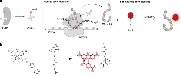
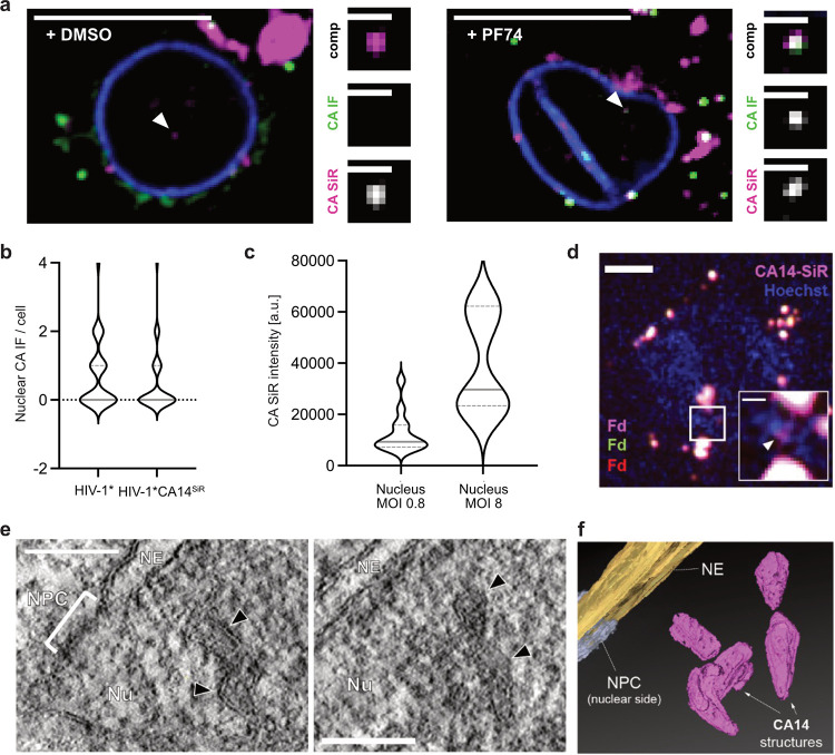
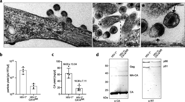
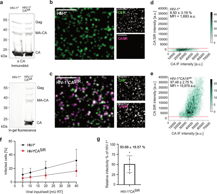
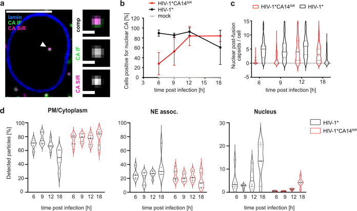
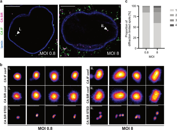
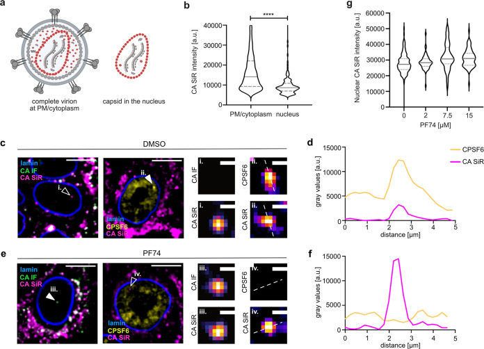
The cone-shaped mature HIV-1 capsid is the main orchestrator of early viral replication. After cytosolic entry, it transports the viral replication complex along microtubules toward the nucleus. While it was initially believed that the reverse transcribed genome is released from the capsid in the cytosol, recent observations indicate that a high amount of capsid protein (CA) remains associated with subviral complexes during import through the nuclear pore complex (NPC). Observation of postentry events via microscopic detection of HIV-1 CA is challenging, since epitope shielding limits immunodetection and the genetic fragility of CA hampers direct labeling approaches. Here, we present a minimally invasive strategy based on genetic code expansion and click chemistry that allows for site-directed fluorescent labeling of HIV-1 CA, while retaining virus morphology and infectivity. Thereby, we could directly visualize virions and subviral complexes using advanced microscopy, including nanoscopy and correlative imaging. Quantification of signal intensities of subviral complexes revealed an amount of CA associated with nuclear complexes in HeLa-derived cells and primary T cells consistent with a complete capsid and showed that treatment with the small molecule inhibitor PF74 did not result in capsid dissociation from nuclear complexes. Cone-shaped objects detected in the nucleus by electron tomography were clearly identified as capsid-derived structures by correlative microscopy. High-resolution imaging revealed dose-dependent clustering of nuclear capsids, suggesting that incoming particles may follow common entry routes. The cone-shaped capsid of HIV-1 has recently been recognized as a master organizer of events from cell entry of the virus to the integration of the viral genome into the host cell DNA. Fluorescent labeling of the capsid is essential to study its role in these dynamic events by microscopy, but viral capsid proteins are extremely challenging targets for the introduction of labels. Here we describe a minimally invasive strategy that allows us to visualize the HIV-1 capsid protein in infected cells by live-cell imaging and superresolution microscopy. Applying this strategy, we confirmed that, contrary to earlier assumptions, an equivalent of a complete capsid can enter the host cell nucleus through nuclear pores. We also observed that entering capsids cluster in the nucleus in a dose-dependent manner, suggesting that they may have followed a common entry route to a site suitable for viral genome release.
成熟的 HIV-1 衣壳呈锥形,是早期病毒复制的主要调控者。衣壳进入细胞质后,沿着微管向核内运输病毒复制复合物。虽然最初人们认为逆转录的基因组在细胞质中从衣壳中释放出来,但最近的观察表明,在通过核孔复合物(NPC)导入时,大量的衣壳蛋白(CA)仍然与亚病毒复合物相关联。通过显微镜观察 HIV-1 CA 的进入后事件具有挑战性,因为表位屏蔽限制了免疫检测,并且 CA 的遗传不稳定性阻碍了直接标记方法。在这里,我们提出了一种基于遗传密码扩展和点击化学的微创策略,该策略允许对 HIV-1 CA 进行定点荧光标记,同时保留病毒形态和感染力。因此,我们可以使用高级显微镜,包括纳米显微镜和相关成像,直接观察病毒粒子和亚病毒复合物。对亚病毒复合物信号强度的定量分析表明,在 HeLa 衍生细胞和原代 T 细胞中与核复合物相关的 CA 数量与完整衣壳一致,并且表明用小分子抑制剂 PF74 处理不会导致衣壳从核复合物中解离。通过电子断层摄影术在核中检测到的锥形物体通过相关显微镜被明确鉴定为衣壳衍生结构。高分辨率成像显示核衣壳的剂量依赖性聚集,表明传入的颗粒可能遵循共同的进入途径。最近,HIV-1 的锥形衣壳被认为是病毒进入细胞到病毒基因组整合到宿主细胞 DNA 过程中各种事件的主要组织者。通过显微镜对衣壳进行荧光标记对于研究其在这些动态事件中的作用至关重要,但病毒衣壳蛋白是引入标记的极具挑战性的目标。在这里,我们描述了一种微创策略,通过活细胞成像和超分辨率显微镜可以使我们观察到感染细胞中的 HIV-1 衣壳蛋白。应用此策略,我们证实与早期假设相反,相当数量的完整衣壳可以通过核孔进入宿主细胞的核。我们还观察到进入的衣壳以剂量依赖性方式在核中聚集,这表明它们可能已经通过了一个共同的进入途径到达了适合释放病毒基因组的部位。