Jorge Rodrigo, Coelho Igor, Viani Gustavo, Vieira Amanda Alexia R, Chahud Fernando, Abud Daniel G, Correa Zelia M
Department of Ophthalmology, Ribeirão Preto Medical School, University of São Paulo, 3900, Bandeirantes Ave, SP, Ribeirão Preto, 14049-900, Brazil.
Department of Medical Images, Hematology and Oncology, Ribeirão Preto Medical School, University of São Paulo, Ribeirão Preto, Brazil.
Int J Retina Vitreous. 2022 Aug 17;8(1):55. doi: 10.1186/s40942-022-00404-1.
Intra-arterial chemotherapy (IAC) has been used to treat multiple cancers including liver metastasis from uveal and cutaneous melanoma but not as primary tumor treatment. We report the compassionate use of chemoreduction with intra-arterial melphalan before ruthenium brachytherapy to salvage an eye with choroidal melanoma.
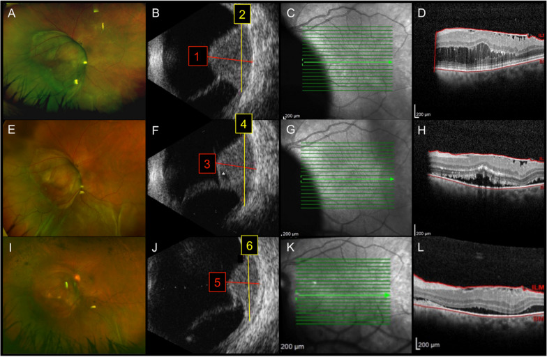
A 61-year-old female patient complained of decreased vision and central-temporal scotoma in OS (left eye) for 1 month. Visual acuity was 20/20 in right eye (OD) and 20/125 OS. Anterior segment examination and intraocular pressure were unremarkable in both eyes, as was fundus examination of the OD. Fundus examination of OS revealed a brown, solid tumor partially obscuring the temporal optic disc margin and extending to the equatorial fundus midzone. Serous retinal detachment was present over the lesion and around it. Ultrasonography revealed a solid choroidal tumor with a largest basal diameter (LBD) of 13.0 mm and thickness of 10.4 mm. The tumor presented acoustic hollowness and a superimposing retinal detachment. After metastatic screening was negative, the patient underwent intra-arterial chemotherapy with melphalan. Three weeks later, her visual acuity was 20/200 and there was noticeable tumor regression to 11.9 mm (LBD) by 7.9 mm (thickness) allowing brachytherapy to be performed. Ten weeks after brachytherapy (13 weeks after IAC), visual acuity was HM due to biopsy-related vitreous hemorrhage (VH). Tumor dimensions were 9.9 (LBD) mm and 6.5 mm (thickness) and PPV was performed to remove VH. Six weeks after PPV (20 weeks after IAC), her visual acuity was 20/200 and further reduction of tumor dimensions was observed: largest basal diameter was 8.9 mm and thickness was 4.9 mm.
This case illustrates the feasibility of combining induction IAC prior to ruthenium brachytherapy for large choroidal melanoma. More studies are warranted to confirm these early preliminary findings.
动脉内化疗(IAC)已被用于治疗多种癌症,包括葡萄膜和皮肤黑色素瘤的肝转移,但未用于原发性肿瘤治疗。我们报告了在钌近距离放射治疗前使用动脉内美法仑进行化疗减容以挽救患有脉络膜黑色素瘤的眼睛的同情用药情况。
一名61岁女性患者主诉左眼视力下降和颞侧中央暗点1个月。右眼视力为20/20,左眼视力为20/125。双眼眼前节检查和眼压均无异常,右眼眼底检查也无异常。左眼眼底检查发现一个棕色实性肿瘤,部分遮挡颞侧视盘边缘并延伸至赤道眼底中区。病变及其周围存在浆液性视网膜脱离。超声检查显示一个实性脉络膜肿瘤,最大基底直径(LBD)为13.0mm,厚度为10.4mm。肿瘤表现为声学空洞和叠加的视网膜脱离。转移筛查为阴性后,患者接受了动脉内美法仑化疗。三周后,她的视力为20/200,肿瘤明显缩小至11.9mm(LBD)×7.9mm(厚度),从而可以进行近距离放射治疗。近距离放射治疗10周后(IAC后13周),由于活检相关的玻璃体积血(VH),视力为眼前手动。肿瘤大小为9.9(LBD)mm×6.5mm(厚度),进行了玻璃体切割术以清除VH。玻璃体切割术后六周(IAC后20周),她的视力为20/200,肿瘤尺寸进一步缩小:最大基底直径为8.9mm,厚度为4.9mm。
本病例说明了在钌近距离放射治疗前联合诱导IAC治疗大型脉络膜黑色素瘤的可行性。需要更多研究来证实这些早期初步发现。