• 文献检索
  • 文档翻译
  • 深度研究
  • 学术资讯
  • Suppr Zotero 插件Zotero 插件
  • 邀请有礼
  • 套餐&价格
  • 历史记录
应用&插件
Suppr Zotero 插件Zotero 插件浏览器插件Mac 客户端Windows 客户端微信小程序
定价
高级版会员购买积分包购买API积分包
服务
文献检索文档翻译深度研究API 文档MCP 服务
关于我们
关于 Suppr公司介绍联系我们用户协议隐私条款
关注我们

Suppr 超能文献

核心技术专利:CN118964589B侵权必究
粤ICP备2023148730 号-1Suppr @ 2026

文献检索

告别复杂PubMed语法,用中文像聊天一样搜索,搜遍4000万医学文献。AI智能推荐,让科研检索更轻松。

立即免费搜索

文件翻译

保留排版,准确专业,支持PDF/Word/PPT等文件格式,支持 12+语言互译。

免费翻译文档

深度研究

AI帮你快速写综述,25分钟生成高质量综述,智能提取关键信息,辅助科研写作。

立即免费体验

免疫组织化学研究 PD-1/PD-L1 通路在皮肤红斑狼疮中的作用。

Immunohistochemical Study of the PD-1/PD-L1 Pathway in Cutaneous Lupus Erythematosus.

机构信息

Department of Dermatology, Venereology and Dermatooncology, Semmelweis University, Budapest, Hungary.

1st Department of Pathology and Experimental Cancer Research, Semmelweis University, Budapest, Hungary.

出版信息

Pathol Oncol Res. 2022 Aug 1;28:1610521. doi: 10.3389/pore.2022.1610521. eCollection 2022.

DOI:10.3389/pore.2022.1610521
PMID:35979531
原文链接:https://pmc.ncbi.nlm.nih.gov/articles/PMC9377145/
Abstract

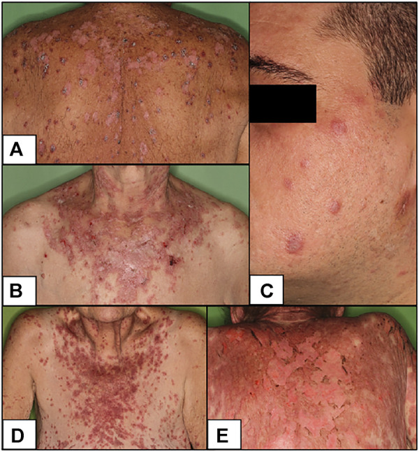
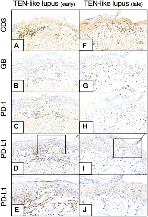
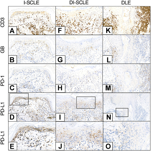
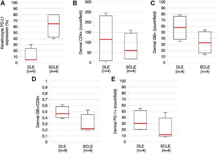
The pathomechanism of various autoimmune diseases is known to be associated with the altered function of programmed cell death 1/programmed cell death ligand 1 (PD-1/PD-L1) axis. We aimed to investigate the role of this pathway and inflammatory cell markers in subtypes of cutaneous lupus erythematosus (CLE): discoid lupus erythematosus (DLE), subacute CLE (SCLE) and toxic epidermal necrolysis (TEN)-like lupus, a hyperacute form of acute CLE (ACLE). Ten skin biopsy samples from 9 patients were analyzed with immunohistochemistry regarding the following markers: CD3, CD4, CD8, Granzyme B, CD123, CD163, PD-1, PD-L1. Our group consisted of 4 SCLE (2 idiopathic (I-SCLE) and 2 PD-1 inhibitor-induced (DI-SCLE)), 4 DLE and 1 TEN-like lupus cases. From the latter patient two consecutive biopsies were obtained 1 week apart. Marker expression patterns were compared through descriptive analysis. Higher median keratinocyte (KC) PD-L1 expression was observed in the SCLE group compared to the DLE group (65% and 5%, respectively). Medians of dermal CD4, Granzyme B (GB), PD-1 positive cell numbers and GB+/CD8 ratio were higher in the DLE group than in the SCLE group. The I-SCLE and DI-SCLE cases showed many similarities, however KC PD-L1 expression and dermal GB positive cell number was higher in the former. The consecutive samples of the TEN-like lupus patient showed an increase by time within the number of infiltrating GB+ cytotoxic T-cells and KC PD-L1 expression (from 22 to 43 and 30%-70%, respectively). Alterations of the PD-1/PD-L1 axis seems to play a role in the pathogenesis of CLE.

摘要

各种自身免疫性疾病的发病机制已知与程序性细胞死亡 1/程序性细胞死亡配体 1(PD-1/PD-L1)轴的功能改变有关。我们旨在研究该途径和炎症细胞标志物在皮肤红斑狼疮(CLE)的亚型中的作用:盘状红斑狼疮(DLE)、亚急性 CLE(SCLE)和毒性表皮坏死松解症(TEN)样狼疮,一种急性 CLE(ACLE)的超急性形式。我们使用免疫组织化学分析了 9 名患者的 10 个皮肤活检样本,分析了以下标志物:CD3、CD4、CD8、颗粒酶 B、CD123、CD163、PD-1、PD-L1。我们的研究小组包括 4 例 SCLE(2 例特发性(I-SCLE)和 2 例 PD-1 抑制剂诱导(DI-SCLE))、4 例 DLE 和 1 例 TEN 样狼疮病例。从后一位患者中连续获得了 2 个间隔 1 周的活检样本。通过描述性分析比较了标志物表达模式。与 DLE 组相比,SCLE 组的角质形成细胞(KC)PD-L1 表达中位数更高(分别为 65%和 5%)。DLE 组的真皮 CD4、颗粒酶 B(GB)、PD-1 阳性细胞数和 GB+/CD8 比值中位数高于 SCLE 组。I-SCLE 和 DI-SCLE 病例有许多相似之处,但前者 KC PD-L1 表达和真皮 GB 阳性细胞数更高。TEN 样狼疮患者的连续样本显示,随着时间的推移,浸润性 GB+细胞毒性 T 细胞和 KC PD-L1 表达的数量增加(分别从 22 到 43 和 30%-70%)。PD-1/PD-L1 轴的改变似乎在 CLE 的发病机制中起作用。

https://cdn.ncbi.nlm.nih.gov/pmc/blobs/fd2b/9377145/d7fc5c107ea5/pore-28-1610521-g004.jpg
https://cdn.ncbi.nlm.nih.gov/pmc/blobs/fd2b/9377145/6b5eae5e4ca1/pore-28-1610521-g001.jpg
https://cdn.ncbi.nlm.nih.gov/pmc/blobs/fd2b/9377145/608b9dde924c/pore-28-1610521-g002.jpg
https://cdn.ncbi.nlm.nih.gov/pmc/blobs/fd2b/9377145/3e5802c60f62/pore-28-1610521-g003.jpg
https://cdn.ncbi.nlm.nih.gov/pmc/blobs/fd2b/9377145/d7fc5c107ea5/pore-28-1610521-g004.jpg

相似文献

1
Immunohistochemical Study of the PD-1/PD-L1 Pathway in Cutaneous Lupus Erythematosus.免疫组织化学研究 PD-1/PD-L1 通路在皮肤红斑狼疮中的作用。
Pathol Oncol Res. 2022 Aug 1;28:1610521. doi: 10.3389/pore.2022.1610521. eCollection 2022.
2
Skin CD4 Trm cells distinguish acute cutaneous lupus erythematosus from localized discoid lupus erythematosus/subacute cutaneous lupus erythematosus and other skin diseases.皮肤 CD4 Trm 细胞可将急性皮肤红斑狼疮与局限性盘状红斑狼疮/亚急性皮肤红斑狼疮和其他皮肤病区分开来。
J Autoimmun. 2022 Apr;128:102811. doi: 10.1016/j.jaut.2022.102811. Epub 2022 Mar 9.
3
The Possible Clinical Significance of a Decreased Serum Level of Soluble PD-L1 in Discoid Lupus Erythematosus, but Not in Subacute Cutaneous Lupus Erythematosus-A Pilot Study.血清可溶性程序性死亡配体1水平降低在盘状红斑狼疮中的可能临床意义,而非亚急性皮肤型红斑狼疮——一项初步研究
J Clin Med. 2023 Aug 30;12(17):5648. doi: 10.3390/jcm12175648.
4
Clinical, histologic, and immunofluorescent distinctions between subacute cutaneous lupus erythematosus and discoid lupus erythematosus.亚急性皮肤型红斑狼疮与盘状红斑狼疮的临床、组织学及免疫荧光鉴别
J Invest Dermatol. 1992 Sep;99(3):251-7. doi: 10.1111/1523-1747.ep12616582.
5
Inflammatory chemokine profiles and their correlations with effector CD4 T cell and regulatory cell subpopulations in cutaneous lupus erythematosus.在红斑狼疮皮损中炎症趋化因子谱及其与效应性 CD4 T 细胞和调节性细胞亚群的相关性。
Cytokine. 2019 Jul;119:95-112. doi: 10.1016/j.cyto.2019.03.010. Epub 2019 Mar 20.
6
B Cell Signatures Distinguish Cutaneous Lupus Erythematosus Subtypes and the Presence of Systemic Disease Activity.B 细胞特征可区分红斑狼疮各亚型及系统性疾病活动度。
Front Immunol. 2021 Nov 19;12:775353. doi: 10.3389/fimmu.2021.775353. eCollection 2021.
7
Characterization of the inflammatory infiltrate and expression of endothelial cell adhesion molecules in lupus erythematosus tumidus.肿胀性红斑狼疮中炎症浸润的特征及内皮细胞黏附分子的表达
Arch Dermatol Res. 2002 Mar;294(1-2):6-13. doi: 10.1007/s00403-001-0286-7.
8
Scarring skin lesions of discoid lupus erythematosus are characterized by high numbers of skin-homing cytotoxic lymphocytes associated with strong expression of the type I interferon-induced protein MxA.盘状红斑狼疮的瘢痕性皮肤病变的特征是大量归巢于皮肤的细胞毒性淋巴细胞,这些细胞与I型干扰素诱导蛋白Mx A的强表达有关。
Br J Dermatol. 2005 Nov;153(5):1011-5. doi: 10.1111/j.1365-2133.2005.06784.x.
9
Upregulation of epidermal surface molecule expression in primary and ultraviolet-induced lesions of lupus erythematosus tumidus.肿胀性红斑狼疮原发性及紫外线诱导性皮损中表皮表面分子表达上调
Br J Dermatol. 2002 May;146(5):801-9. doi: 10.1046/j.1365-2133.2002.04693.x.
10
Subacute cutaneous lupus erythematosus and discoid lupus erythematosus. Comparative histopathologic findings.亚急性皮肤型红斑狼疮和盘状红斑狼疮。组织病理学比较结果。
Arch Dermatol. 1984 Mar;120(3):332-7.

引用本文的文献

1
The Possible Clinical Significance of a Decreased Serum Level of Soluble PD-L1 in Discoid Lupus Erythematosus, but Not in Subacute Cutaneous Lupus Erythematosus-A Pilot Study.血清可溶性程序性死亡配体1水平降低在盘状红斑狼疮中的可能临床意义,而非亚急性皮肤型红斑狼疮——一项初步研究
J Clin Med. 2023 Aug 30;12(17):5648. doi: 10.3390/jcm12175648.

本文引用的文献

1
Subacute cutaneous lupus erythematosus induced by PD-1 Inhibitor therapy: two case reports and literature review.PD-1抑制剂治疗诱发的亚急性皮肤型红斑狼疮:两例病例报告及文献综述
Australas J Dermatol. 2021 May;62(2):e347-e349. doi: 10.1111/ajd.13538. Epub 2021 Jan 23.
2
Systematic Review: Monoclonal Antibody-Induced Subacute Cutaneous Lupus Erythematosus.系统评价:单克隆抗体诱导的亚急性皮肤型红斑狼疮。
Drugs R D. 2020 Dec;20(4):319-330. doi: 10.1007/s40268-020-00320-5.
3
De novo subacute cutaneous lupus erythematosus-like eruptions in the setting of programmed death-1 or programmed death ligand-1 inhibitor therapy: clinicopathological correlation.
程序性死亡受体-1 或程序性死亡配体-1 抑制剂治疗中出现新发亚急性皮肤狼疮样疹:临床病理相关性。
Clin Exp Dermatol. 2021 Mar;46(2):328-337. doi: 10.1111/ced.14449. Epub 2020 Oct 14.
4
Systemic lupus erythematosus onset in patient receiving anti-PD1 treatment with pembrolizumab: a case report.接受派姆单抗抗PD1治疗的患者发生系统性红斑狼疮:一例报告
Rheumatology (Oxford). 2021 Feb 1;60(2):e39-e40. doi: 10.1093/rheumatology/keaa389.
5
Immune-related adverse events of checkpoint inhibitors.检查点抑制剂的免疫相关不良反应。
Nat Rev Dis Primers. 2020 May 7;6(1):38. doi: 10.1038/s41572-020-0160-6.
6
Evaluation of PD-L1, PD-L2, PD-1 and cytotoxic immune response in oral lichen planus.口腔扁平苔藓中PD-L1、PD-L2、PD-1及细胞毒性免疫反应的评估
Oral Dis. 2020 Sep;26(6):1246-1254. doi: 10.1111/odi.13344. Epub 2020 Apr 16.
7
PD-1H (VISTA)-mediated suppression of autoimmunity in systemic and cutaneous lupus erythematosus.PD-1H(VISTA)介导的系统性红斑狼疮和皮肤红斑狼疮自身免疫抑制作用。
Sci Transl Med. 2019 Dec 11;11(522). doi: 10.1126/scitranslmed.aax1159.
8
Cutaneous lupus erythematosus induced by drugs - novel insights.药物诱导的皮肤狼疮红斑 - 新的见解。
Expert Rev Clin Pharmacol. 2020 Jan;13(1):35-42. doi: 10.1080/17512433.2020.1698290. Epub 2019 Dec 2.
9
Are there distinct clinical and pathological features distinguishing idiopathic from drug-induced subacute cutaneous lupus erythematosus? A European retrospective multicenter study.特发性与药物诱导的亚急性皮肤型狼疮是否具有独特的临床和病理特征?一项欧洲回顾性多中心研究。
J Am Acad Dermatol. 2019 Aug;81(2):403-411. doi: 10.1016/j.jaad.2019.02.009. Epub 2019 Feb 11.
10
Cutaneous lupus associated with pembrolizumab therapy for advanced melanoma: a report of three cases.帕博利珠单抗治疗晚期黑色素瘤相关皮肤狼疮:三例报告。
Melanoma Res. 2019 Jun;29(3):338-341. doi: 10.1097/CMR.0000000000000587.