Perez Alexandre, Lenoir Vincent, Lombardi Tommaso
Unit of Oral Surgery and Implantology, Division of Oral and Maxillofacial Surgery, Department of Surgery, University of Geneva & University Hospitals of Geneva, 1205 Geneva, Switzerland.
Division of Radiology, Diagnostic Department, Geneva University Hospitals, University of Geneva, 1205 Geneva, Switzerland.
Diagnostics (Basel). 2022 Aug 19;12(8):2006. doi: 10.3390/diagnostics12082006.
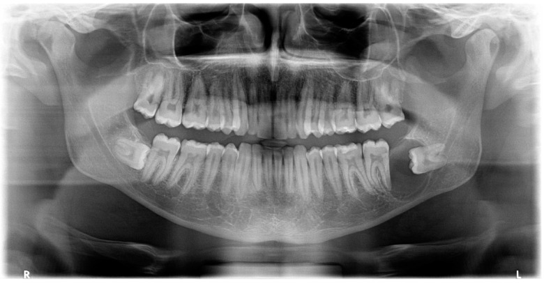
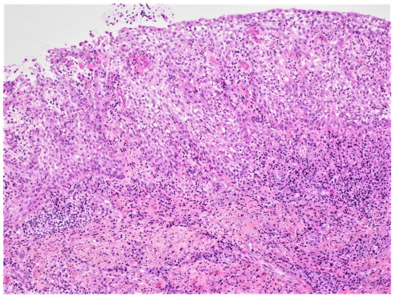
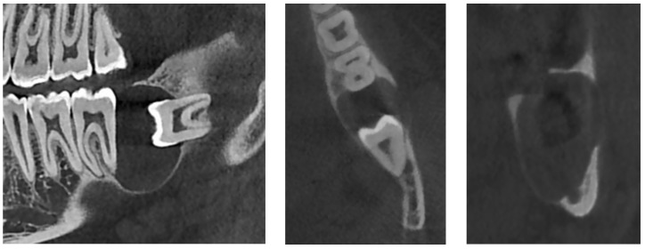
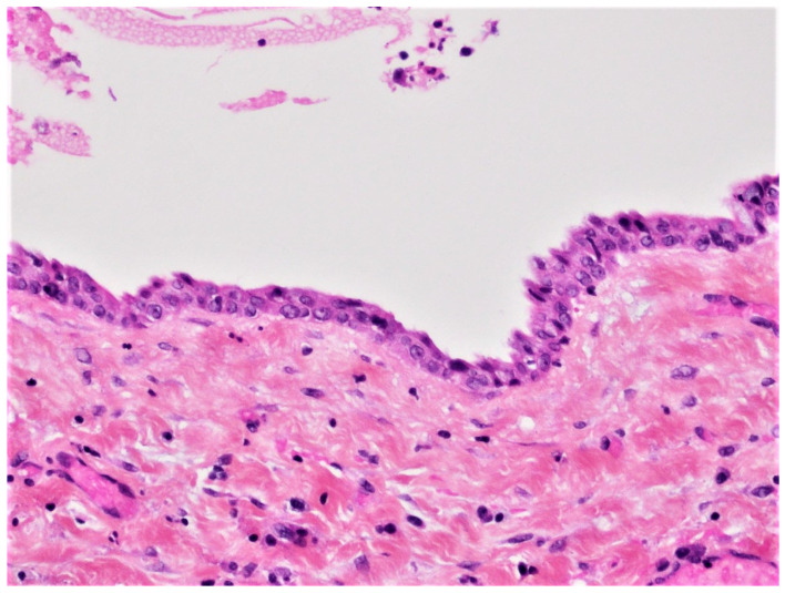
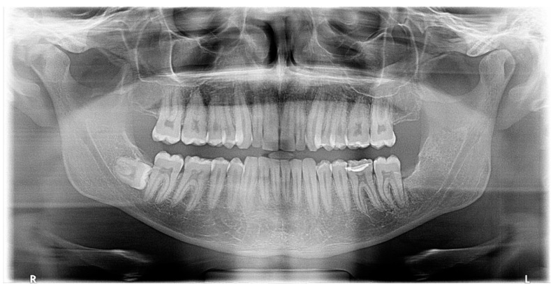
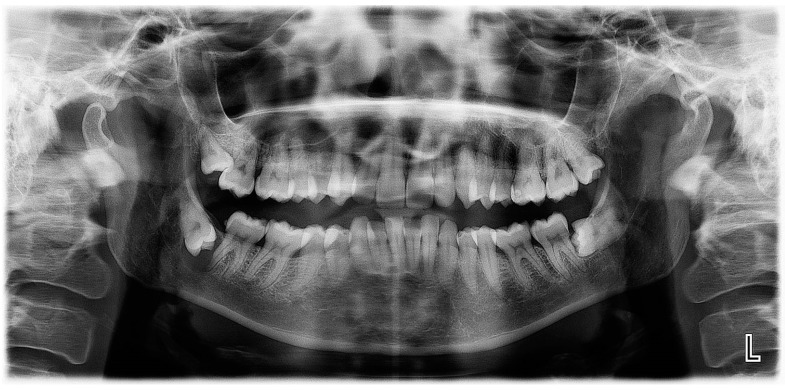
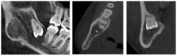
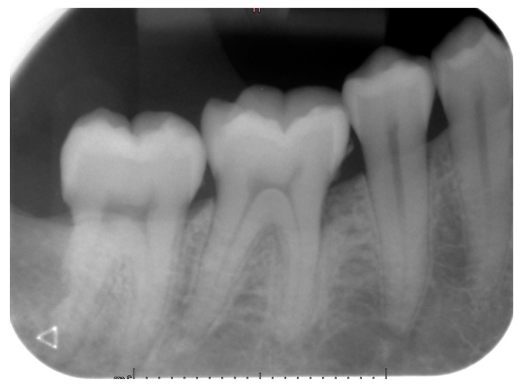
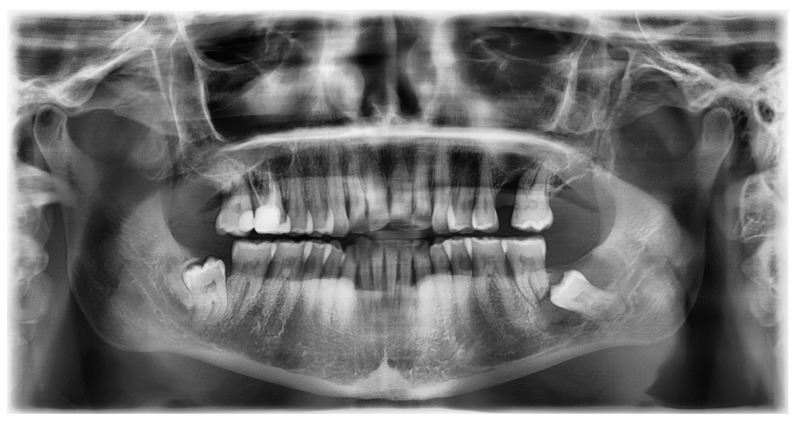
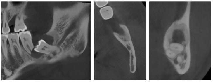
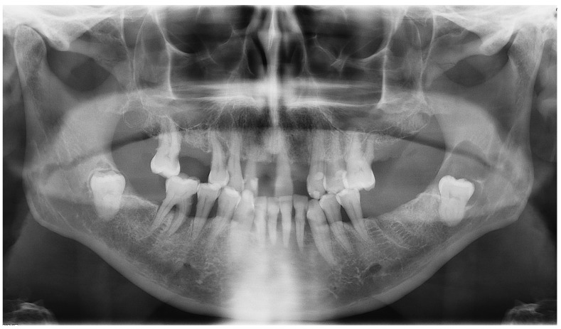
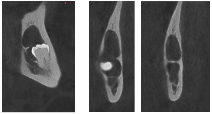
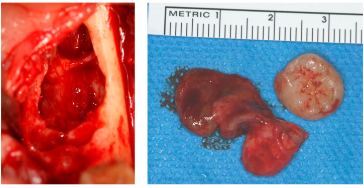
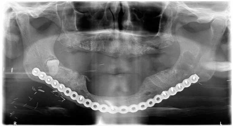
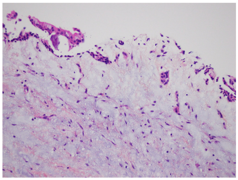
Dentigerous cyst is an odontogenic developmental cyst arising from the pericoronal tissue of an impacted tooth, and that may exhibit various radiological aspects. The aim of this article is to present four cases of histologically confirmed mandibular dentigerous cysts to highlight diverse radiological presentations: one of classical appearance (well-limited unilocular radiolucent lesion surrounding the crown) and three which have shown radiological peculiarities (one cyst displacing the adjacent tooth, with bone but no root resorption, one cyst presenting hallmarks of infection and one multilocular cyst with thin septa). Such radiologic diversity may, on occasion, suggest a clinical aggressive lesion such as an odontogenic keratocyst or ameloblastoma. The diagnosis of dentigerous cyst requires a thorough evaluation of the clinical presentation and accurate radiological studies.
含牙囊肿是一种牙源性发育性囊肿,起源于阻生牙的冠周组织,可能呈现出各种影像学表现。本文旨在介绍4例经组织学证实的下颌含牙囊肿病例,以突出其多样的影像学表现:1例为典型表现(围绕牙冠的界限清晰的单房透射性病变),3例表现出影像学特点(1例囊肿使相邻牙齿移位,有骨质但无牙根吸收;1例囊肿有感染特征;1例为具有薄分隔的多房囊肿)。这种影像学的多样性有时可能提示临床侵袭性病变,如牙源性角化囊肿或成釉细胞瘤。含牙囊肿的诊断需要对临床表现进行全面评估并进行准确的影像学检查。