Iwata Eiji, Kusumoto Junya, Hasegawa Takumi, Tachibana Akira, Akashi Masaya
Oral and Maxillofacial Surgery, Kakogawa Central City Hospital, Kakogawa, JPN.
Oral and Maxillofacial Surgery, Kobe University Hospital, Kobe, JPN.
Cureus. 2024 Oct 3;16(10):e70791. doi: 10.7759/cureus.70791. eCollection 2024 Oct.
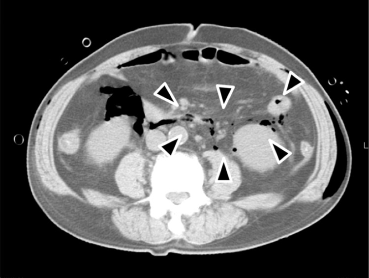
Mandibular wisdom teeth can occasionally cause infections, which can progress to severe deep neck infections (DNIs) including deep neck abscesses or necrotizing soft tissue infections, which are fatal. This study aimed to identify the radiographic characteristics of mandibular wisdom teeth that developed severe DNIs.
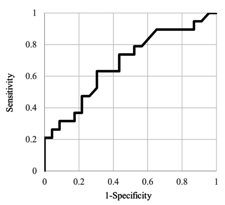
This study included patients who were admitted for the treatment of severe mandibular wisdom tooth infection between July 2012 and June 2024 at a single center. Patient characteristics, clinical data, and radiographic findings were analyzed and compared between the severe DNI group and mild DNI group including patients with cellulitis or superficial abscess. < 0.05 was considered significant.
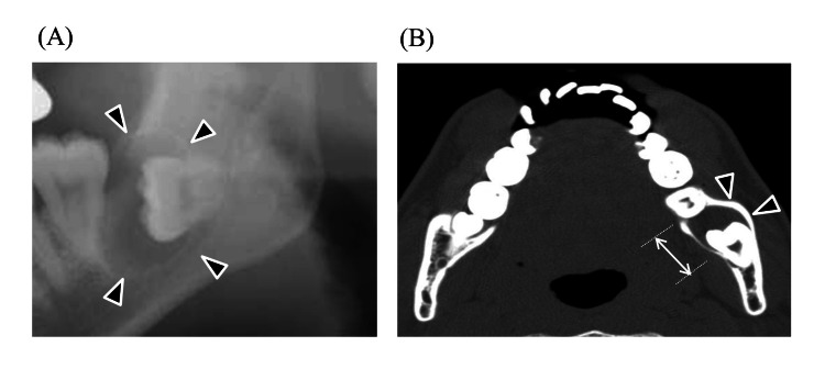
Nineteen of 42 patients (45.2%) were included in the severe DNI group. The multivariate analysis showed that the highest odds ratio (OR) was for the presence of a radicular cyst (OR=17.7), followed by the presence of a dentigerous cyst (OR =14.5). The most common mandibular wisdom tooth with a dentigerous cyst in patients with severe DNIs was inverted according to Winter's classification and type IIIC in the Pell and Gregory classification.
Radiographic characteristics associated with severe DNIs included the presence of radicular and dentigerous cysts in the mandibular wisdom teeth. Especially in dentigerous cysts, deeply impacted teeth should be taken attention.
下颌智齿偶尔会引发感染,进而发展为严重的深部颈部感染(DNIs),包括深部颈部脓肿或坏死性软组织感染,这些感染可能会致命。本研究旨在确定发生严重DNIs的下颌智齿的影像学特征。
本研究纳入了2012年7月至2024年6月期间在单一中心因严重下颌智齿感染入院治疗的患者。对严重DNI组与包括蜂窝织炎或浅表脓肿患者的轻度DNI组之间的患者特征、临床数据和影像学表现进行分析和比较。P < 0.05被认为具有统计学意义。
42例患者中有19例(45.2%)被纳入严重DNI组。多因素分析显示,根囊肿的存在的比值比(OR)最高(OR = 17.7),其次是含牙囊肿的存在(OR = 14.5)。根据温特分类法,严重DNIs患者中含牙囊肿最常见的下颌智齿呈倒置,在佩尔和格雷戈里分类中为IIIC型。
与严重DNIs相关的影像学特征包括下颌智齿中存在根囊肿和含牙囊肿。特别是在含牙囊肿中,对于深度阻生的牙齿应予以关注。