• 文献检索
  • 文档翻译
  • 深度研究
  • 学术资讯
  • Suppr Zotero 插件Zotero 插件
  • 邀请有礼
  • 套餐&价格
  • 历史记录
应用&插件
Suppr Zotero 插件Zotero 插件浏览器插件Mac 客户端Windows 客户端微信小程序
定价
高级版会员购买积分包购买API积分包
服务
文献检索文档翻译深度研究API 文档MCP 服务
关于我们
关于 Suppr公司介绍联系我们用户协议隐私条款
关注我们

Suppr 超能文献

核心技术专利:CN118964589B侵权必究
粤ICP备2023148730 号-1Suppr @ 2026

文献检索

告别复杂PubMed语法,用中文像聊天一样搜索,搜遍4000万医学文献。AI智能推荐,让科研检索更轻松。

立即免费搜索

文件翻译

保留排版,准确专业,支持PDF/Word/PPT等文件格式,支持 12+语言互译。

免费翻译文档

深度研究

AI帮你快速写综述,25分钟生成高质量综述,智能提取关键信息,辅助科研写作。

立即免费体验

功能成像和解剖成像模态在相关转移性嗜铬细胞瘤和副神经节瘤中的表现

Performances of Functional and Anatomic Imaging Modalities in Related Metastatic Pheochromocytoma and Paraganglioma.

作者信息

Patel Mayank, Jha Abhishek, Ling Alexander, Chen Clara C, Millo Corina, Kuo Mickey J M, Nazari Matthew A, Talvacchio Sara, Charles Kailah, Miettinen Markku, Del Rivero Jaydira, Chen Alice P, Nilubol Naris, Lin Frank I, Civelek Ali Cahid, Taïeb David, Carrasquillo Jorge A, Pacak Karel

机构信息

Section on Medical Neuroendocrinology, Eunice Kennedy Shriver National Institute of Child Health and Human Development, National Institutes of Health, Bethesda, MD 20814, USA.

Radiology and Imaging Sciences, Warren Grant Magnuson Clinical Center, National Institutes of Health, Bethesda, MD 20814, USA.

出版信息

Cancers (Basel). 2022 Aug 11;14(16):3886. doi: 10.3390/cancers14163886.

DOI:10.3390/cancers14163886
PMID:36010880
原文链接:https://pmc.ncbi.nlm.nih.gov/articles/PMC9406057/
Abstract

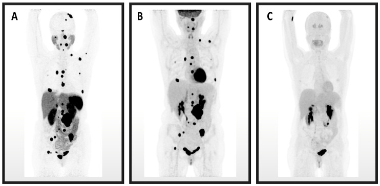
The study identifies the importance of positron emission tomographic (PET) and anatomic imaging modalities and their individual performances in detecting succinate dehydrogenase A (-related metastatic pheochromocytoma and paraganglioma (PPGL). The detection rates of PET modalities-Ga-DOTATATE, F-FDG, and F-FDOPA-along with the combination of computed tomography (CT) and magnetic resonance imaging (MRI) are compared in a cohort of 11 patients with metastatic PPGL in the setting of a germline mutation. The imaging detection performances were evaluated at three levels: overall lesions, anatomic regions, and a patient-by-patient basis. Ga-DOTATATE PET demonstrated a lesion-based detection rate of 88.6% [95% confidence interval (CI), 84.3-92.5%], while F-FDG, F-FDOPA, and CT/MRI showed detection rates of 82.9% (CI, 78.0-87.1%), 39.8% (CI, 30.2-50.2%), and 58.2% (CI, 52.0-64.1%), respectively. The study found that Ga-DOTATATE best detects lesions in a subset of patients with related metastatic PPGL. However, F-FDG did detect more lesions in the liver, mediastinum, and abdomen/pelvis anatomic regions, showing the importance of a combined approach using both PET modalities in evaluating -related PPGL.

摘要

该研究确定了正电子发射断层扫描(PET)和解剖成像模式的重要性及其在检测琥珀酸脱氢酶A相关转移性嗜铬细胞瘤和副神经节瘤(PPGL)中的个体表现。在一组11例患有种系突变的转移性PPGL患者中,比较了PET模式(镓-多柔比星、氟代脱氧葡萄糖和氟代多巴)以及计算机断层扫描(CT)和磁共振成像(MRI)联合使用的检测率。在三个层面评估成像检测性能:整体病变、解剖区域和逐个患者层面。镓-多柔比星PET显示基于病变的检测率为88.6%[95%置信区间(CI),84.3-92.5%],而氟代脱氧葡萄糖、氟代多巴和CT/MRI的检测率分别为82.9%(CI,78.0-87.1%)、39.8%(CI,30.2-50.2%)和58.2%(CI,52.0-64.1%)。该研究发现,镓-多柔比星在相关转移性PPGL患者亚组中对病变的检测效果最佳。然而,氟代脱氧葡萄糖确实在肝脏、纵隔和腹部/盆腔解剖区域检测到更多病变,这表明在评估相关PPGL时联合使用两种PET模式的重要性。

https://cdn.ncbi.nlm.nih.gov/pmc/blobs/5a23/9406057/aded4fd80a13/cancers-14-03886-g001.jpg

相似文献

1
Performances of Functional and Anatomic Imaging Modalities in Related Metastatic Pheochromocytoma and Paraganglioma.功能成像和解剖成像模态在相关转移性嗜铬细胞瘤和副神经节瘤中的表现
Cancers (Basel). 2022 Aug 11;14(16):3886. doi: 10.3390/cancers14163886.
2
Superiority of Ga-DOTATATE over F-FDG and anatomic imaging in the detection of succinate dehydrogenase mutation (SDHx )-related pheochromocytoma and paraganglioma in the pediatric population.镓-DOTATATE 比 F-FDG 和解剖成像在儿童人群中检测琥珀酸脱氢酶突变(SDHx)相关嗜铬细胞瘤和副神经节瘤方面更具优势。
Eur J Nucl Med Mol Imaging. 2018 May;45(5):787-797. doi: 10.1007/s00259-017-3896-9. Epub 2017 Dec 4.
3
Diagnostic performance of [Ga]DOTATATE PET/CT, [F]FDG PET/CT, MRI of the spine, and whole-body diagnostic CT and MRI in the detection of spinal bone metastases associated with pheochromocytoma and paraganglioma.用于诊断与嗜铬细胞瘤和副神经节瘤相关的脊柱骨转移的[Ga]DOTATATE PET/CT、[F]FDG PET/CT、脊柱 MRI 以及全身诊断性 CT 和 MRI 的诊断性能。
Eur Radiol. 2024 Oct;34(10):6488-6498. doi: 10.1007/s00330-024-10652-4. Epub 2024 Apr 16.
4
Superiority of [68Ga]-DOTATATE PET/CT to Other Functional Imaging Modalities in the Localization of SDHB-Associated Metastatic Pheochromocytoma and Paraganglioma.[68Ga]-DOTATATE PET/CT在SDHB相关转移性嗜铬细胞瘤和副神经节瘤定位方面相对于其他功能成像模态的优越性。
Clin Cancer Res. 2015 Sep 1;21(17):3888-95. doi: 10.1158/1078-0432.CCR-14-2751. Epub 2015 Apr 14.
5
PET/CT comparing (68)Ga-DOTATATE and other radiopharmaceuticals and in comparison with CT/MRI for the localization of sporadic metastatic pheochromocytoma and paraganglioma.PET/CT比较(68)镓-多柔比星和其他放射性药物,并与CT/MRI比较用于散发性转移性嗜铬细胞瘤和副神经节瘤的定位。
Eur J Nucl Med Mol Imaging. 2016 Sep;43(10):1784-91. doi: 10.1007/s00259-016-3357-x. Epub 2016 Mar 21.
6
The utility of Ga-DOTATATE PET/CT in localizing primary/metastatic pheochromocytoma and paraganglioma in children and adolescents - a single-center experience.镓-多他特(Ga-DOTATATE)PET/CT在定位儿童和青少年原发性/转移性嗜铬细胞瘤和副神经节瘤中的应用——单中心经验
J Pediatr Endocrinol Metab. 2020 Nov 12;34(1):109-119. doi: 10.1515/jpem-2020-0354. Print 2021 Jan 27.
7
Clinical, Diagnostic, and Treatment Characteristics of -Related Metastatic Pheochromocytoma and Paraganglioma.与[具体内容缺失]相关的转移性嗜铬细胞瘤和副神经节瘤的临床、诊断及治疗特征
Front Oncol. 2019 Feb 22;9:53. doi: 10.3389/fonc.2019.00053. eCollection 2019.
8
68Ga-DOTATATE PET/CT in the Localization of Head and Neck Paragangliomas Compared with Other Functional Imaging Modalities and CT/MRI.与其他功能成像方式及CT/MRI相比,68Ga-DOTATATE PET/CT在头颈部副神经节瘤定位中的应用
J Nucl Med. 2016 Feb;57(2):186-91. doi: 10.2967/jnumed.115.161018. Epub 2015 Nov 12.
9
Diagnostic Performance of (68)Ga-DOTATATE PET/CT, (18)F-FDG PET/CT and (131)I-MIBG Scintigraphy in Mapping Metastatic Pheochromocytoma and Paraganglioma.(68)镓- DOTATATE PET/CT、(18)氟- FDG PET/CT及(131)碘- MIBG闪烁扫描术在转移性嗜铬细胞瘤和副神经节瘤定位中的诊断效能
Nucl Med Mol Imaging. 2015 Jun;49(2):143-51. doi: 10.1007/s13139-015-0331-7. Epub 2015 Apr 8.
10
The Utility of Ga-DOTATATE PET/CT in Localizing Primary/Metastatic Pheochromocytoma and Paraganglioma: Asian Indian Experience.镓[68Ga] DOTATATE PET/CT在定位原发性/转移性嗜铬细胞瘤和副神经节瘤中的应用:亚洲印度人的经验
Indian J Endocrinol Metab. 2021 Sep-Oct;25(5):410-417. doi: 10.4103/ijem.ijem_307_21. Epub 2022 Jan 12.

引用本文的文献

1
Evaluating the diagnostic accuracy of Tc-labeled somatostatin receptor imaging for suspected pheochromocytomas and paragangliomas.评估锝标记的生长抑素受体显像对疑似嗜铬细胞瘤和副神经节瘤的诊断准确性。
Sci Rep. 2025 Jul 28;15(1):27493. doi: 10.1038/s41598-025-12794-x.
2
Case Series: Variants in Four Patients with Metastatic Pheochromocytoma.病例系列:4 例转移性嗜铬细胞瘤患者的变异情况。
Front Endocrinol (Lausanne). 2024 Sep 16;15:1399847. doi: 10.3389/fendo.2024.1399847. eCollection 2024.
3
Diagnostic performance of [Ga]DOTATATE PET/CT, [F]FDG PET/CT, MRI of the spine, and whole-body diagnostic CT and MRI in the detection of spinal bone metastases associated with pheochromocytoma and paraganglioma.

本文引用的文献

1
Imaging of Pheochromocytoma and Paraganglioma.《嗜铬细胞瘤和副神经节瘤的影像学表现》
J Nucl Med. 2021 Aug 1;62(8):1033-1042. doi: 10.2967/jnumed.120.259689.
2
Personalized Management of Pheochromocytoma and Paraganglioma.《嗜铬细胞瘤和副神经节瘤的个体化管理》
Endocr Rev. 2022 Mar 9;43(2):199-239. doi: 10.1210/endrev/bnab019.
3
International consensus on initial screening and follow-up of asymptomatic SDHx mutation carriers.国际上对无症状 SDHx 突变携带者的初步筛查和随访达成共识。
用于诊断与嗜铬细胞瘤和副神经节瘤相关的脊柱骨转移的[Ga]DOTATATE PET/CT、[F]FDG PET/CT、脊柱 MRI 以及全身诊断性 CT 和 MRI 的诊断性能。
Eur Radiol. 2024 Oct;34(10):6488-6498. doi: 10.1007/s00330-024-10652-4. Epub 2024 Apr 16.
4
Imaging of Pheochromocytomas and Paragangliomas.《嗜铬细胞瘤和副神经节瘤的影像学表现》
Endocr Rev. 2024 May 7;45(3):414-434. doi: 10.1210/endrev/bnae001.
5
Synchronous carotid body and anterior mediastinal paraganglioma revealed by F-fluorodeoxyglucose and Ga-DOTA(0)-Tyr(3)-octreotate positron emission tomography-computed tomography.F-氟脱氧葡萄糖和Ga-DOTA(0)-Tyr(3)-奥曲肽正电子发射断层扫描-计算机断层扫描显示的同步颈动脉体瘤和前纵隔副神经节瘤
Quant Imaging Med Surg. 2023 Jun 1;13(6):4007-4014. doi: 10.21037/qims-22-1018. Epub 2023 Apr 4.
Nat Rev Endocrinol. 2021 Jul;17(7):435-444. doi: 10.1038/s41574-021-00492-3. Epub 2021 May 21.
4
Emerging Treatments for Advanced/Metastatic Pheochromocytoma and Paraganglioma.晚期/转移性嗜铬细胞瘤和副神经节瘤的新兴治疗方法。
Curr Treat Options Oncol. 2020 Aug 29;21(11):85. doi: 10.1007/s11864-020-00787-z.
5
Molecular imaging and radionuclide therapy of pheochromocytoma and paraganglioma in the era of genomic characterization of disease subgroups.疾病亚组基因组特征时代的嗜铬细胞瘤和副神经节瘤的分子成像和放射性核素治疗。
Endocr Relat Cancer. 2019 Nov;26(11):R627-R652. doi: 10.1530/ERC-19-0165.
6
European Association of Nuclear Medicine Practice Guideline/Society of Nuclear Medicine and Molecular Imaging Procedure Standard 2019 for radionuclide imaging of phaeochromocytoma and paraganglioma.欧洲核医学协会实践指南/核医学与分子影像学会2019年嗜铬细胞瘤和副神经节瘤放射性核素成像程序标准
Eur J Nucl Med Mol Imaging. 2019 Sep;46(10):2112-2137. doi: 10.1007/s00259-019-04398-1. Epub 2019 Jun 29.
7
Eruption of Metastatic Paraganglioma After Successful Therapy with Lu/Y-DOTATOC and Lu-DOTATATE.经镥/钇-奥曲肽和镥-奥曲肽成功治疗后转移性副神经节瘤的复发
Nucl Med Mol Imaging. 2019 Jun;53(3):223-230. doi: 10.1007/s13139-019-00579-w. Epub 2019 Feb 22.
8
Clinical, Diagnostic, and Treatment Characteristics of -Related Metastatic Pheochromocytoma and Paraganglioma.与[具体内容缺失]相关的转移性嗜铬细胞瘤和副神经节瘤的临床、诊断及治疗特征
Front Oncol. 2019 Feb 22;9:53. doi: 10.3389/fonc.2019.00053. eCollection 2019.
9
Update of Pheochromocytoma Syndromes: Genetics, Biochemical Evaluation, and Imaging.嗜铬细胞瘤综合征的最新进展:遗传学、生化评估与影像学
Front Endocrinol (Lausanne). 2018 Nov 27;9:515. doi: 10.3389/fendo.2018.00515. eCollection 2018.
10
Bayesian approach to determining penetrance of pathogenic SDH variants.贝叶斯方法在确定致病性 SDH 变异体外显率中的应用。
J Med Genet. 2018 Nov;55(11):729-734. doi: 10.1136/jmedgenet-2018-105427. Epub 2018 Sep 10.