Section on Medical Neuroendocrinology, Eunice Kennedy Shriver National Institute of Child Health and Human Development, National Institutes of Health, Bethesda, MD, United States.
Medical Genetics Branch, National Human Genome Research Institute, National Institutes of Health, Bethesda, MD, United States.
Front Endocrinol (Lausanne). 2024 Sep 16;15:1399847. doi: 10.3389/fendo.2024.1399847. eCollection 2024.
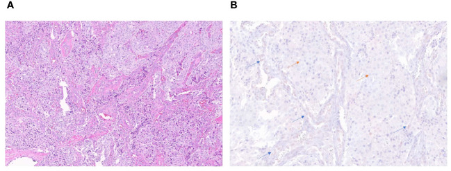
Few reports have highlighted the rare presence of somatic variants in clinically aggressive, metastatic pheochromocytoma/paraganglioma (PCC/PGL); however, none have addressed detailed clinical presentation (including biochemistry and imaging) and management of these patients. Here, we address these clinical features and management based on four PCC patients with somatic variants from our National Institutes of Health PCC/PGL cohort. A total of 192 patients underwent exome sequencing (germline, somatic, or both), and four males were found to have somatic variants (with additional somatic and oncogenic variants in patients 2 and 4, respectively). Per-lesion and per-patient comparisons were performed among functional imaging scans performed at the NIH. Biochemical phenotype and response to systemic treatment were evaluated. This mini-series supports prior studies showing aggressive/metastatic PCC in patients with somatic variants, as all developed widespread metastatic disease. All four PCC patients presented with noradrenergic biochemical phenotype, and some with significant elevation in 3-methoxytyramine. F-FDOPA PET/CT was found to be the superior functional imaging modality, with 100% lesion detection rate when compared to that of Ga-DOTATATE, F-FDG, F-FDA, and I-MIBG scans. While patients did not respond to chemotherapy or tyrosine kinase inhibitors, they responded to targeted radiotherapy using high-specific-activity I-MIBG (Azedra) or Lu-DOTATATE (Lutathera).
鲜有报道强调体细胞变异在临床上侵袭性强、转移性强的嗜铬细胞瘤/副神经节瘤(PCC/PGL)中的罕见存在;然而,目前尚无研究涉及这些患者的详细临床表现(包括生化和影像学)和管理。在此,我们根据来自美国国立卫生研究院 PCC/PGL 队列的 4 名患有体细胞变异的 PCC 患者,介绍这些临床特征和管理方法。共有 192 名患者接受了外显子组测序(种系、体细胞或两者),发现 4 名男性存在体细胞变异(患者 2 和 4 分别存在额外的体细胞和致癌变异)。在 NIH 进行的功能影像学扫描中对病变和患者进行了比较。评估了生化表型和全身治疗的反应。这个迷你系列支持了先前的研究,表明患有体细胞变异的患者存在侵袭性/转移性 PCC,因为所有患者均发展为广泛转移的疾病。所有 4 名 PCC 患者均表现为去甲肾上腺素能生化表型,有些患者的 3-甲氧基酪胺显著升高。与 Ga-DOTATATE、F-FDG、F-FDA 和 I-MIBG 扫描相比,F-FDOPA PET/CT 被发现是更好的功能影像学检查方法,其病变检出率为 100%。虽然患者对化疗或酪氨酸激酶抑制剂无反应,但他们对使用高比活度 I-MIBG(Azedra)或 Lu-DOTATATE(Lutathera)的靶向放疗有反应。