• 文献检索
  • 文档翻译
  • 深度研究
  • 学术资讯
  • Suppr Zotero 插件Zotero 插件
  • 邀请有礼
  • 套餐&价格
  • 历史记录
应用&插件
Suppr Zotero 插件Zotero 插件浏览器插件Mac 客户端Windows 客户端微信小程序
定价
高级版会员购买积分包购买API积分包
服务
文献检索文档翻译深度研究API 文档MCP 服务
关于我们
关于 Suppr公司介绍联系我们用户协议隐私条款
关注我们

Suppr 超能文献

核心技术专利:CN118964589B侵权必究
粤ICP备2023148730 号-1Suppr @ 2026

文献检索

告别复杂PubMed语法,用中文像聊天一样搜索,搜遍4000万医学文献。AI智能推荐,让科研检索更轻松。

立即免费搜索

文件翻译

保留排版,准确专业,支持PDF/Word/PPT等文件格式,支持 12+语言互译。

免费翻译文档

深度研究

AI帮你快速写综述,25分钟生成高质量综述,智能提取关键信息,辅助科研写作。

立即免费体验

18-FDG PET-CT影像组学特征能否预测不可切除或无法切除食管癌的局部无进展生存期?

Could 18-FDG PET-CT Radiomic Features Predict the Locoregional Progression-Free Survival in Inoperable or Unresectable Oesophageal Cancer?

作者信息

De Bari Berardino, Lefevre Loriane, Henriques Julie, Gatta Roberto, Falcoz Antoine, Mathieu Pierre, Borg Christophe, Dinapoli Nicola, Boulahdour Hatem, Boldrini Luca, Valentini Vincenzo, Vernerey Dewi

机构信息

Radiation Oncology Department, Neuchâtel Hospital Network, CH-2300 La Chaux-de-Fonds, Switzerland.

Radiation Oncology Department, University Hospital of Besançon, F-25000 Besancon, France.

出版信息

Cancers (Basel). 2022 Aug 22;14(16):4043. doi: 10.3390/cancers14164043.

DOI:10.3390/cancers14164043
PMID:36011035
原文链接:https://pmc.ncbi.nlm.nih.gov/articles/PMC9406583/
Abstract

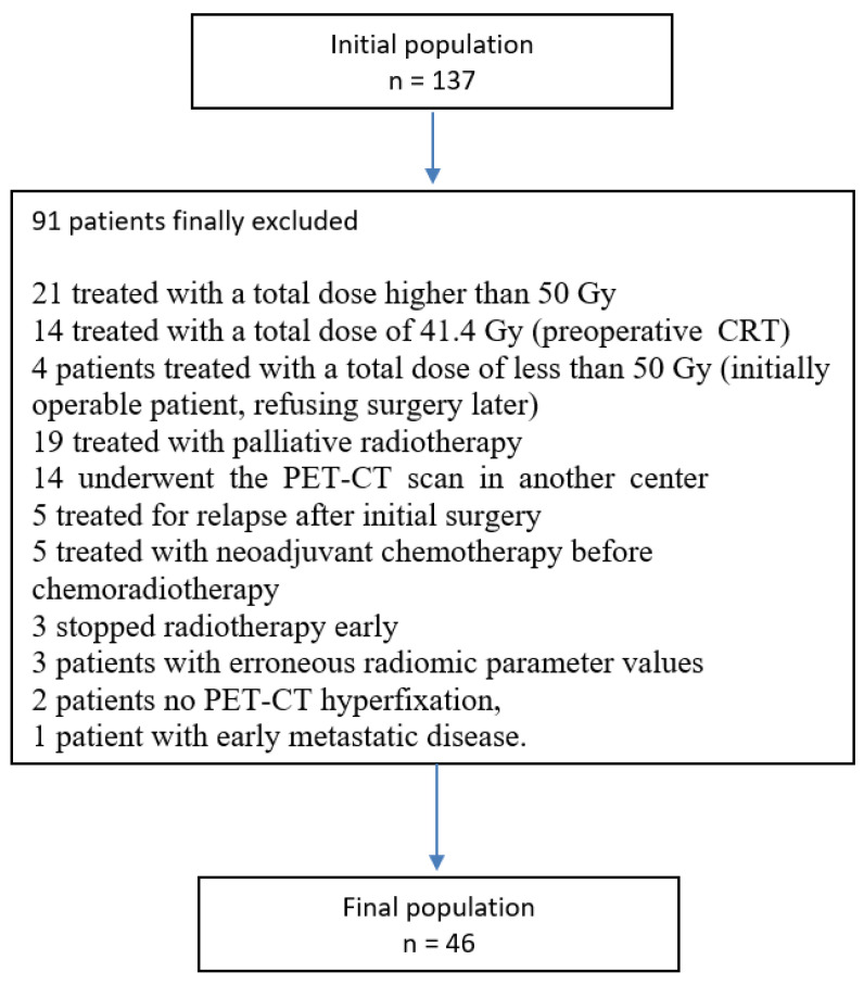
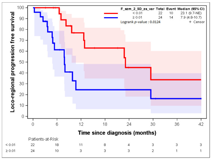
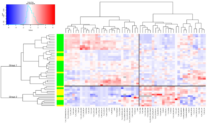
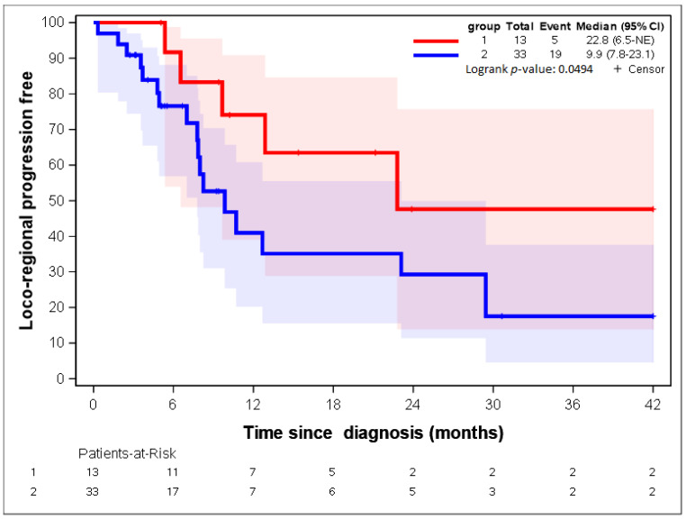
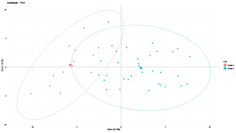
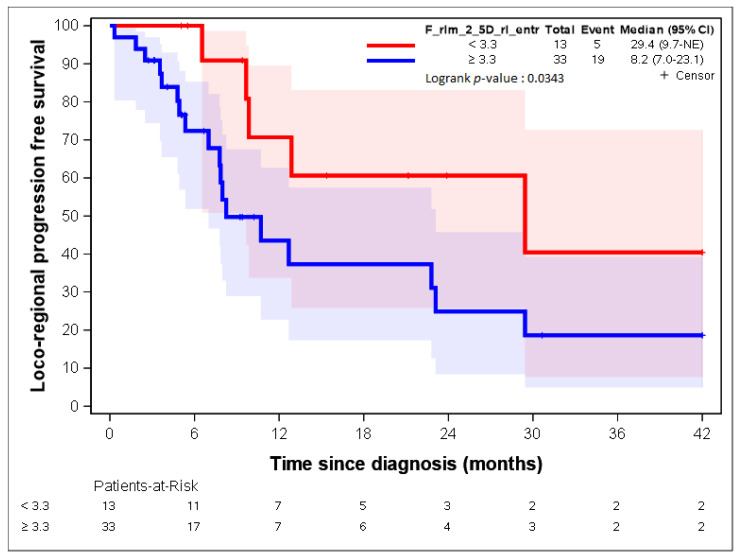
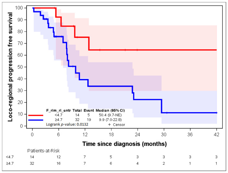
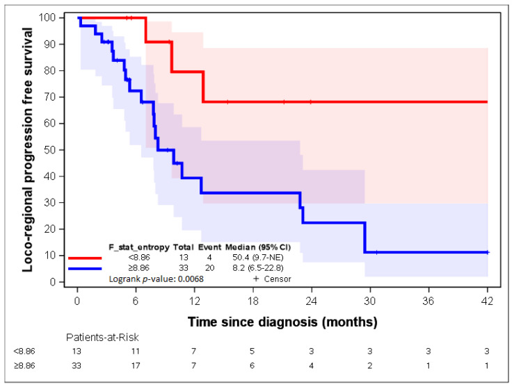
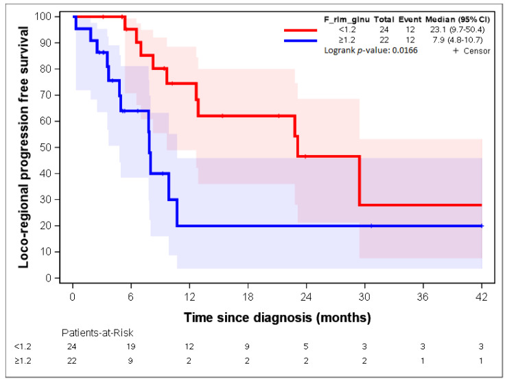
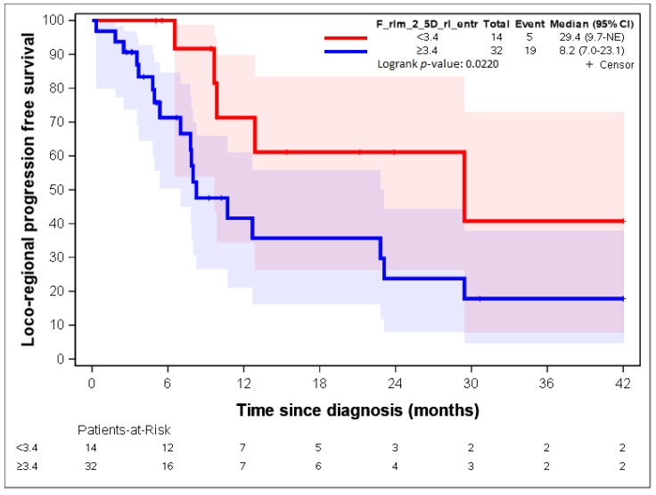
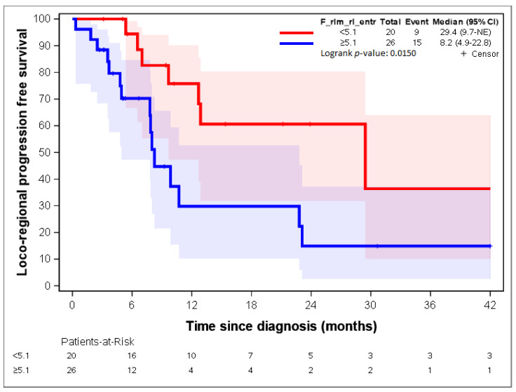
Background: We evaluated the value of pre-treatment positron-emission tomography−computed tomography (PET-CT)-based radiomic features in predicting the locoregional progression-free survival (LR-PFS) of patients with inoperable or unresectable oesophageal cancer. Material and Methods: Forty-six patients were included and 230 radiomic parameters were extracted. After a principal component analysis (PCA), we identified the more robust radiomic parameters, and we used them to develop a heatmap. Finally, we correlated these radiomic features with LR-PFS. Results: The median follow-up time was 17 months. The two-year LR-PFS and PFS rates were 35.9% (95% CI: 18.9−53.3) and 21.6% (95%CI: 10.0−36.2), respectively. After the correlation analysis, we identified 55 radiomic parameters that were included in the heatmap. According to the results of the hierarchical clustering, we identified two groups of patients presenting statistically different median LR-PFSs (22.8 months vs. 9.9 months; HR = 2.64; 95% CI 0.97−7.15; p = 0.0573). We also identified two radiomic features (“F_rlm_rl_entr_per” and “F_rlm_2_5D_rl_entr”) significantly associated with LR-PFS. Patients expressing a “F_rlm_2_5D_rl_entr” of <3.3 had a better median LR- PFS (29.4 months vs. 8.2 months; p = 0.0343). Patients presenting a “F_rlm_rl_entr_per” of <4.7 had a better median LR-PFS (50.4 months vs. 9.9 months; p = 0.0132). Conclusion: We identified two radiomic signatures associated with a lower risk of locoregional relapse after CRT.

摘要

背景

我们评估了基于治疗前正电子发射断层扫描-计算机断层扫描(PET-CT)的放射组学特征在预测不可手术或无法切除的食管癌患者局部区域无进展生存期(LR-PFS)方面的价值。

材料与方法

纳入46例患者,提取230个放射组学参数。经过主成分分析(PCA)后,我们确定了更稳健的放射组学参数,并用于制作热图。最后,我们将这些放射组学特征与LR-PFS进行关联。

结果

中位随访时间为17个月。两年LR-PFS率和PFS率分别为35.9%(95%CI:18.9−53.3)和21.6%(95%CI:10.0−36.2)。经过相关性分析,我们确定了55个纳入热图的放射组学参数。根据层次聚类结果,我们确定了两组患者,其LR-PFS中位数在统计学上存在差异(22.8个月对9.9个月;HR = 2.64;95%CI 0.97−7.15;p = 0.0573)。我们还确定了两个与LR-PFS显著相关的放射组学特征(“F_rlm_rl_entr_per”和“F_rlm_2_5D_rl_entr”)。“F_rlm_2_5D_rl_entr”<3.3的患者具有更好的LR-PFS中位数(29.4个月对8.2个月;p = 0.0343)。“F_rlm_rl_entr_per”<4.7的患者具有更好的LR-PFS中位数(50.4个月对9.9个月;p = 0.0132)。

结论

我们确定了两个与CRT后局部区域复发风险较低相关的放射组学特征。

https://cdn.ncbi.nlm.nih.gov/pmc/blobs/7f15/9406583/44fafaef7f22/cancers-14-04043-g011.jpg
https://cdn.ncbi.nlm.nih.gov/pmc/blobs/7f15/9406583/46ed2b0f0efd/cancers-14-04043-g001.jpg
https://cdn.ncbi.nlm.nih.gov/pmc/blobs/7f15/9406583/150607b96a5e/cancers-14-04043-g002.jpg
https://cdn.ncbi.nlm.nih.gov/pmc/blobs/7f15/9406583/b770019f5802/cancers-14-04043-g003.jpg
https://cdn.ncbi.nlm.nih.gov/pmc/blobs/7f15/9406583/170bb169ed54/cancers-14-04043-g004.jpg
https://cdn.ncbi.nlm.nih.gov/pmc/blobs/7f15/9406583/281ca1eba81f/cancers-14-04043-g005.jpg
https://cdn.ncbi.nlm.nih.gov/pmc/blobs/7f15/9406583/30e17f1c7be1/cancers-14-04043-g006.jpg
https://cdn.ncbi.nlm.nih.gov/pmc/blobs/7f15/9406583/171dd30e3f35/cancers-14-04043-g007.jpg
https://cdn.ncbi.nlm.nih.gov/pmc/blobs/7f15/9406583/41b6dc28c552/cancers-14-04043-g008.jpg
https://cdn.ncbi.nlm.nih.gov/pmc/blobs/7f15/9406583/bb9ca3011597/cancers-14-04043-g009.jpg
https://cdn.ncbi.nlm.nih.gov/pmc/blobs/7f15/9406583/ef4bc7c87383/cancers-14-04043-g010.jpg
https://cdn.ncbi.nlm.nih.gov/pmc/blobs/7f15/9406583/44fafaef7f22/cancers-14-04043-g011.jpg

相似文献

1
Could 18-FDG PET-CT Radiomic Features Predict the Locoregional Progression-Free Survival in Inoperable or Unresectable Oesophageal Cancer?18-FDG PET-CT影像组学特征能否预测不可切除或无法切除食管癌的局部无进展生存期?
Cancers (Basel). 2022 Aug 22;14(16):4043. doi: 10.3390/cancers14164043.
2
Development and validation of an [F]FDG-PET/CT radiomic model for predicting progression-free survival for patients with stage II - III thoracic esophageal squamous cell carcinoma who are treated with definitive chemoradiotherapy.用于预测接受根治性放化疗的II - III期胸段食管鳞状细胞癌患者无进展生存期的[F]FDG-PET/CT放射组学模型的开发与验证
Acta Oncol. 2023 Feb;62(2):159-165. doi: 10.1080/0284186X.2023.2178859. Epub 2023 Feb 15.
3
Prediction of outcome in anal squamous cell carcinoma using radiomic feature analysis of pre-treatment FDG PET-CT.基于治疗前 FDG PET-CT 影像组学特征分析预测肛门鳞癌患者的预后。
Eur J Nucl Med Mol Imaging. 2019 Dec;46(13):2790-2799. doi: 10.1007/s00259-019-04495-1. Epub 2019 Sep 4.
4
Pretreatment CT and F-FDG PET-based radiomic model predicting pathological complete response and loco-regional control following neoadjuvant chemoradiation in oesophageal cancer.基于预处理 CT 和 F-FDG PET 的放射组学模型预测食管癌新辅助放化疗后病理完全缓解和局部区域控制
J Med Imaging Radiat Oncol. 2021 Feb;65(1):102-111. doi: 10.1111/1754-9485.13128. Epub 2020 Dec 1.
5
[ F]-FDG PET radiomic model as prognostic biomarker in diffuse large B-cell lymphoma.[F]-氟代脱氧葡萄糖正电子发射断层扫描放射组学模型作为弥漫性大B细胞淋巴瘤的预后生物标志物
Hematol Oncol. 2023 Oct;41(4):674-682. doi: 10.1002/hon.3171. Epub 2023 May 20.
6
Radiomics in vulvar cancer: first clinical experience using F-FDG PET/CT images.外阴癌的影像组学:使用F-FDG PET/CT图像的首次临床经验
J Nucl Med. 2018 Jul 20;60(2):199-206. doi: 10.2967/jnumed.118.215889.
7
Prediction of local recurrence and distant metastasis using radiomics analysis of pretreatment nasopharyngeal [18F]FDG PET/CT images.基于治疗前鼻咽部 [18F]FDG PET/CT 图像的放射组学分析预测局部复发和远处转移。
Ann Nucl Med. 2021 Apr;35(4):458-468. doi: 10.1007/s12149-021-01585-9. Epub 2021 Feb 4.
8
The role of FDG PET/CT radiomics in the prediction of pathological response to neoadjuvant treatment in patients with esophageal cancer.氟代脱氧葡萄糖正电子发射断层扫描/计算机断层扫描(FDG PET/CT)影像组学在预测食管癌患者新辅助治疗的病理反应中的作用。
Rep Pract Oncol Radiother. 2024 Jun 6;29(2):211-218. doi: 10.5603/rpor.99906. eCollection 2024.
9
Radiomic features of glucose metabolism enable prediction of outcome in mantle cell lymphoma.基于葡萄糖代谢的放射组学特征可预测套细胞淋巴瘤的预后。
Eur J Nucl Med Mol Imaging. 2019 Dec;46(13):2760-2769. doi: 10.1007/s00259-019-04420-6. Epub 2019 Jul 8.
10
Is PET Radiomics Useful to Predict Pathologic Tumor Response and Prognosis in Locally Advanced Cervical Cancer?PET 放射组学在预测局部晚期宫颈癌的病理肿瘤反应和预后方面是否有用?
J Nucl Med. 2024 Jun 3;65(6):962-970. doi: 10.2967/jnumed.123.267044.

引用本文的文献

1
Expression of Concern: De Bari et al. Could 18-FDG PET-CT Radiomic Features Predict the Locoregional Progression-Free Survival in Inoperable or Unresectable Oesophageal Cancer? 2022, , 4043.关注声明:德巴里等人。18-FDG PET-CT放射组学特征能否预测不可切除或无法切除的食管癌的局部无进展生存期?2022年,,4043。
Cancers (Basel). 2025 Apr 23;17(9):1400. doi: 10.3390/cancers17091400.
2
Integrating F-FDG PET/CT radiomics and body composition for enhanced prognostic assessment in patients with esophageal cancer.将 F-FDG PET/CT 影像组学与身体成分相结合,以增强食管癌患者的预后评估。
BMC Cancer. 2024 Nov 14;24(1):1402. doi: 10.1186/s12885-024-13157-x.

本文引用的文献

1
18F-FDG PET Radiomics as Predictor of Treatment Response in Oesophageal Cancer: A Systematic Review and Meta-Analysis.18F-FDG PET 影像组学作为食管癌治疗反应的预测指标:一项系统评价与Meta分析
Front Oncol. 2022 Mar 15;12:861638. doi: 10.3389/fonc.2022.861638. eCollection 2022.
2
The Image Biomarker Standardization Initiative: Standardized Quantitative Radiomics for High-Throughput Image-based Phenotyping.影像生物标志物标准化倡议:高通量基于影像表型的标准化定量放射组学。
Radiology. 2020 May;295(2):328-338. doi: 10.1148/radiol.2020191145. Epub 2020 Mar 10.
3
Exploring technical issues in personalized medicine: NSCLC survival prediction by quantitative image analysis-usefulness of density correction of volumetric CT data.
探讨个性化医学中的技术问题:通过定量图像分析预测 NSCLC 患者的生存情况——容积 CT 数据密度校正的作用。
Radiol Med. 2020 Jul;125(7):625-635. doi: 10.1007/s11547-020-01157-3. Epub 2020 Mar 3.
4
Integrating radiomics into holomics for personalised oncology: from algorithms to bedside.将放射组学纳入整体组学以进行个体化肿瘤学:从算法到床边。
Eur Radiol Exp. 2020 Feb 7;4(1):11. doi: 10.1186/s41747-019-0143-0.
5
Combining the radiomic features and traditional parameters of F-FDG PET with clinical profiles to improve prognostic stratification in patients with esophageal squamous cell carcinoma treated with neoadjuvant chemoradiotherapy and surgery.将 F-FDG PET 的放射组学特征和传统参数与临床特征相结合,以改善接受新辅助放化疗和手术治疗的食管鳞癌患者的预后分层。
Ann Nucl Med. 2019 Sep;33(9):657-670. doi: 10.1007/s12149-019-01380-7. Epub 2019 Jun 19.
6
Clinical complete response after chemoradiotherapy for carcinoma of thoracic esophagus: Is esophagectomy always necessary? A systematic review and meta-analysis.放化疗后胸段食管癌的临床完全缓解:是否一定要行手术切除?系统评价和荟萃分析。
Thorac Cancer. 2018 Dec;9(12):1638-1647. doi: 10.1111/1759-7714.12874. Epub 2018 Oct 1.
7
The Role of PET-Based Radiomic Features in Predicting Local Control of Esophageal Cancer Treated with Concurrent Chemoradiotherapy.基于 PET 的放射组学特征在预测同步放化疗治疗食管癌局部控制中的作用。
Sci Rep. 2018 Jul 2;8(1):9902. doi: 10.1038/s41598-018-28243-x.
8
Chemoradiotherapy versus chemoradiotherapy plus surgery for esophageal cancer.食管癌的化疗放疗与化疗放疗联合手术治疗对比
Cochrane Database Syst Rev. 2017 Aug 22;8(8):CD010511. doi: 10.1002/14651858.CD010511.pub2.
9
Radiomics Features of Multiparametric MRI as Novel Prognostic Factors in Advanced Nasopharyngeal Carcinoma.多参数 MRI 放射组学特征作为晚期鼻咽癌的新型预后因素。
Clin Cancer Res. 2017 Aug 1;23(15):4259-4269. doi: 10.1158/1078-0432.CCR-16-2910. Epub 2017 Mar 9.
10
Points of View: Heat maps.观点:热图。
Nat Methods. 2012 Mar;9(3):213. doi: 10.1038/nmeth.1902.