Daminova Amina G, Rogov Alexey M, Rassabina Anna E, Beckett Richard P, Minibayeva Farida V
Kazan Institute of Biochemistry and Biophysics, FRC Kazan Scientific Center, Russian Academy of Sciences, P.O. Box 261, 420111 Kazan, Russia.
Interdisciplinary Center for Analytical Microscopy, Kazan (Volga Region) Federal University, 420018 Kazan, Russia.
J Fungi (Basel). 2022 Jul 28;8(8):791. doi: 10.3390/jof8080791.
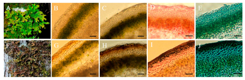
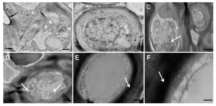
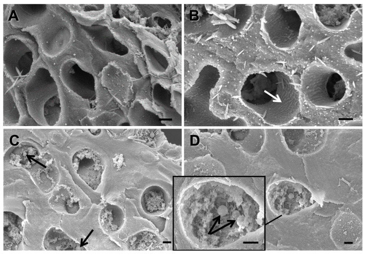
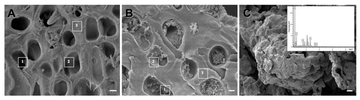
Lichens often grow in microhabitats where they experience severe abiotic stresses. Some species respond to high UV radiation by synthesizing dark brown melanic pigments in the upper cortex. However, unlike the melanized structures of non-lichenized fungi, the morphology of the melanic layer in lichens remains unstudied. Here, we analyzed the morphology, ultrastructure, and elemental composition of the melanized layer in UV-exposed thalli of the lichen (L.) Hoffm. Using light microscopy, we detected a pigmented layer sensitive to staining with 3,4-L-dihydroxyphenylalanine, a precursor of eumelanin, in the upper cortex of melanized thalli. Analysis of cross-sections of melanized thalli using scanning electron microscopy revealed that melanin-like granules are deposited into the hyphal lumens. Melanized thalli also possessed thicker hyphal cell walls compared to pale thalli. Energy-dispersive X-ray spectroscopy analysis of the elemental composition of the hyphal walls and extracted melanin indicated that the type of melanin synthesized by is eumelanin. Transmission electron microscopy was used to show that during melanization melanosome-like dark vesicles are transported to the cell surface and secreted into the cell walls of the fungal hyphae. Results from this study provide new insights into the effects of melanin synthesis on the microstructure of lichen thalli.
地衣通常生长在经历严重非生物胁迫的微生境中。一些物种通过在上皮层合成深褐色的黑色素来应对高紫外线辐射。然而,与非地衣化真菌的黑化结构不同,地衣中黑化层的形态仍未得到研究。在此,我们分析了地衣(L.)霍夫曼暴露于紫外线的叶状体中黑化层的形态、超微结构和元素组成。使用光学显微镜,我们在黑化叶状体的上层皮层中检测到一层对真黑素前体3,4-L-二羟基苯丙氨酸染色敏感的色素层。使用扫描电子显微镜对黑化叶状体的横截面进行分析,结果显示类黑色素颗粒沉积在菌丝腔内。与浅色叶状体相比,黑化叶状体的菌丝细胞壁也更厚。对菌丝壁和提取的黑色素的元素组成进行能量色散X射线光谱分析表明,合成的黑色素类型为真黑素。透射电子显微镜用于显示在黑化过程中,类黑素体的深色囊泡被运输到细胞表面并分泌到真菌菌丝的细胞壁中。这项研究的结果为黑色素合成对地衣叶状体微观结构的影响提供了新的见解。