Biomedical Engineering and Imaging Institute, Icahn School of Medicine at Mount Sinai, New York, USA.
Department of Diagnostic, Molecular and Interventional Radiology, Icahn School of Medicine at Mount Sinai, New York, USA.
NMR Biomed. 2023 Jan;36(1):e4823. doi: 10.1002/nbm.4823. Epub 2022 Sep 23.
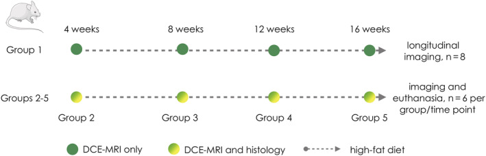
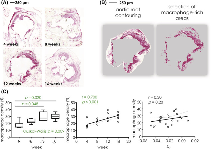
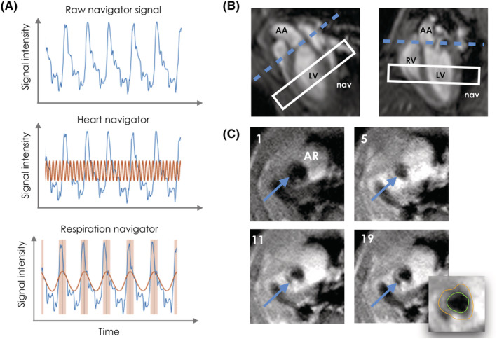
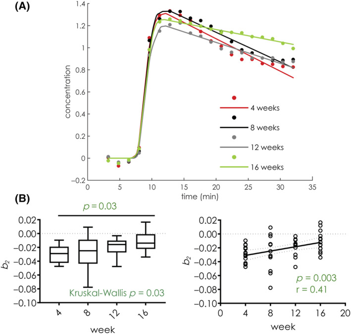
High-risk atherosclerotic plaques are characterized by active inflammation and abundant leaky microvessels. We present a self-gated, dynamic contrast-enhanced magnetic resonance imaging (DCE-MRI) acquisition with compressed sensing reconstruction and apply it to assess longitudinal changes in endothelial permeability in the aortic root of Apoe atherosclerotic mice during natural disease progression. Twenty-four, 8-week-old, female Apoe mice were divided into four groups (n = 6 each) and imaged with self-gated DCE-MRI at 4, 8, 12, and 16 weeks after high-fat diet initiation, and then euthanized for CD68 immunohistochemistry for macrophages. Eight additional mice were kept on a high-fat diet and imaged longitudinally at the same time points. Aortic-root pseudo-concentration curves were analyzed using a validated piecewise linear model. Contrast agent wash-in and washout slopes (b and b ) were measured as surrogates of aortic root endothelial permeability and compared with macrophage density by immunohistochemistry. b , indicating contrast agent washout, was significantly higher in mice kept on an high-fat diet for longer periods of time (p = 0.03). Group comparison revealed significant differences between mice on a high-fat diet for 4 versus 16 weeks (p = 0.03). Macrophage density also significantly increased with diet duration (p = 0.009). Spearman correlation between b from DCE-MRI and macrophage density indicated a weak relationship between the two parameters (r = 0.28, p = 0.20). Validated piecewise linear modeling of the DCE-MRI data showed that the aortic root contrast agent washout rate is significantly different during disease progression. Further development of this technique from a single-slice to a 3D acquisition may enable better investigation of the relationship between in vivo imaging of endothelial permeability and atherosclerotic plaques' genetic, molecular, and cellular makeup in this important model of disease.
高危动脉粥样硬化斑块的特征是活跃的炎症和丰富的渗漏微血管。我们提出了一种自门控、动态对比增强磁共振成像(DCE-MRI)采集方法,并应用该方法评估载脂蛋白 E (Apoe)动脉粥样硬化小鼠主动脉根部内皮通透性在自然疾病进展过程中的纵向变化。24 只 8 周龄雌性 Apoe 小鼠被分为 4 组(每组 6 只),在高脂饮食开始后 4、8、12 和 16 周时进行自门控 DCE-MRI 成像,然后处死进行巨噬细胞 CD68 免疫组化。另外 8 只小鼠继续高脂饮食,并在相同时间点进行纵向成像。使用验证的分段线性模型分析主动脉根部伪浓度曲线。测量对比剂的洗脱斜率(b 和 b )作为主动脉根部内皮通透性的替代物,并与免疫组化检测的巨噬细胞密度进行比较。b ,表示对比剂洗脱,在高脂饮食时间较长的小鼠中显著升高(p=0.03)。组间比较显示,高脂饮食 4 周与 16 周的小鼠之间存在显著差异(p=0.03)。巨噬细胞密度也随着饮食时间的延长而显著增加(p=0.009)。DCE-MRI 的 b 值与巨噬细胞密度之间的 Spearman 相关性表明这两个参数之间存在弱相关性(r=0.28,p=0.20)。DCE-MRI 数据的分段线性模型验证表明,在疾病进展过程中,主动脉根部对比剂的洗脱率存在显著差异。从单层面到 3D 采集的这种技术的进一步发展可能使我们能够更好地研究这个重要疾病模型中内皮通透性的体内成像与动脉粥样硬化斑块的遗传、分子和细胞成分之间的关系。