Kunugiza Yasuo, Tanaka Takehiro, Hirota Ryuichiro, Kakunaga Shigeki, Okamoto Yasunori, Tsuji Shigeyoshi
Department of Orthopaedics, Japan Community Health Care Organization (JCHO), Hoshigaoka Medical Center, Hoshigaoka 4-8-1, Osaka, Japan.
Department of Radiology, Japan Community Health Care Organization (JCHO), Hoshigaoka Medical Center, Osaka, Japan.
Radiol Case Rep. 2022 Aug 17;17(10):3987-3991. doi: 10.1016/j.radcr.2022.07.070. eCollection 2022 Oct.
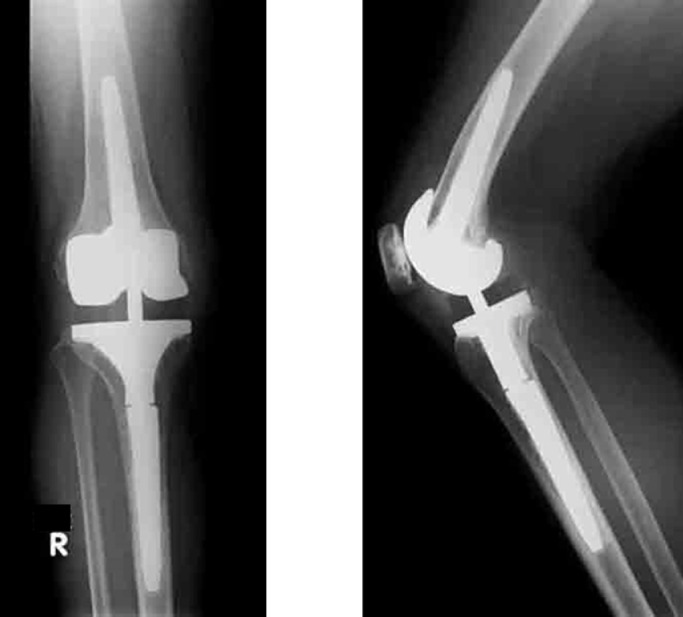
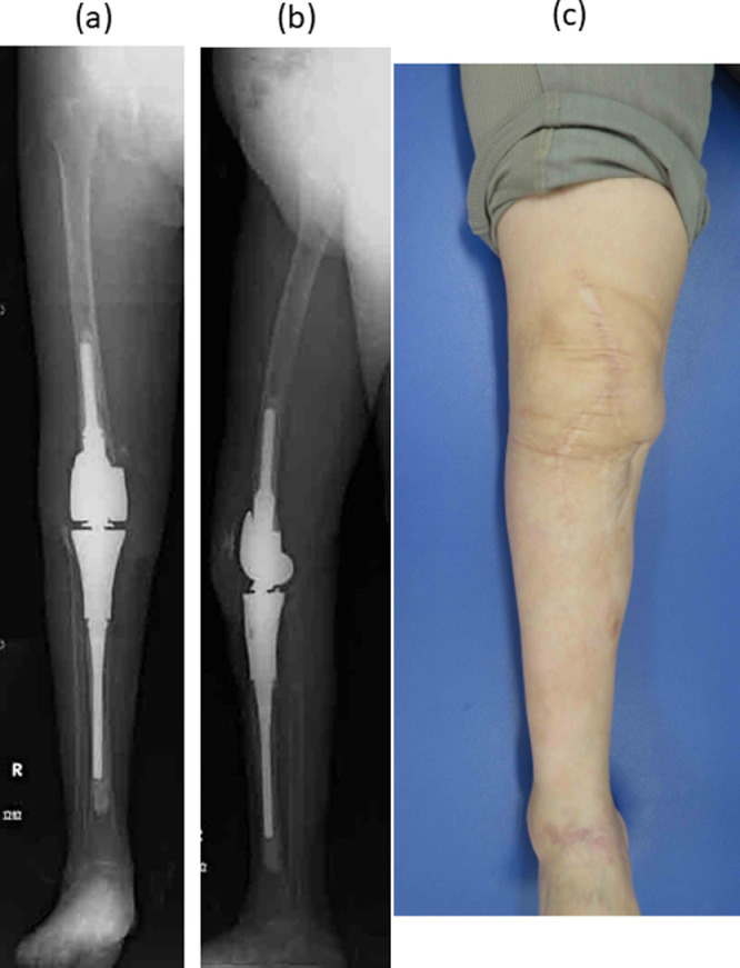
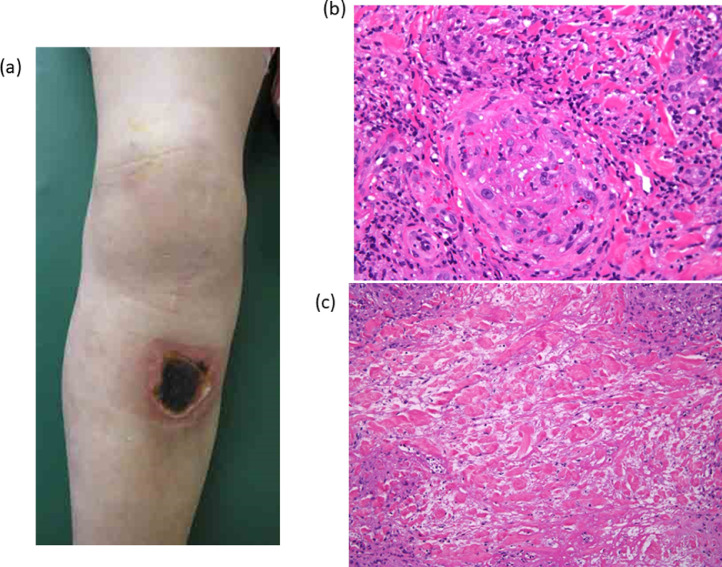
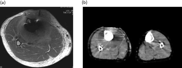
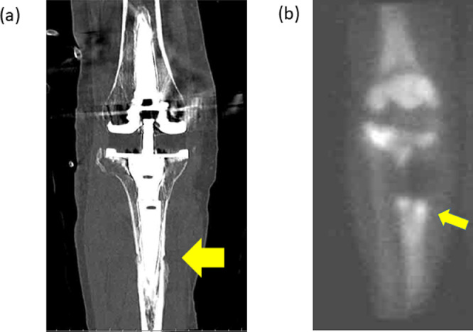
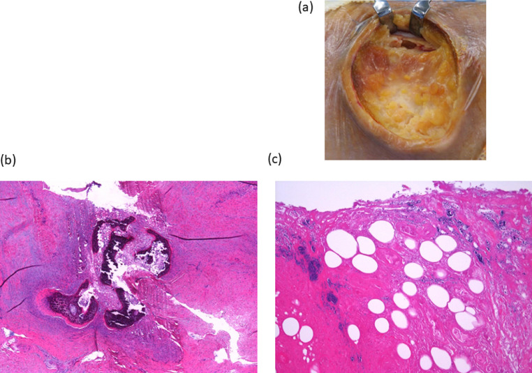
We report the case of a 71-year-old woman with a skin ulcer derived from an abscess around the tibia. The abscess resulted in periprosthetic joint infection and osteomyelitis 11 years after total knee arthroplasty. The first symptom was a skin ulcer of the lower leg. Magnetic resonance imaging revealed a circumferential mass around the proximal tibia. A skin biopsy taken around the ulcer showed thrombosis and degenerated collagen. Contrast-enhanced computed tomography showed a circumferential mass around the proximal tibia with ring enhancement. Biopsies of the skin ulcer and circumferential mass showed an abscess caused by and methicillin-resistant . We conducted debridement of the abscess, a gastrocnemius flap and split-thickness skin grafting and a 2-stage revision of the total knee component with a hinged prosthesis. Two years later, the infection did not reoccur and the patient can walk without a cane. This case is unique as abscess around proximal tibia caused necrotic skin ulcer and appearance of abscess was fibrous and different from typical bacterial abscesses containing pus or fluid. Contrast-enhanced computed tomography was effective for differentiation of the pathological condition.
我们报告了一例71岁女性患者,其皮肤溃疡源自胫骨周围的脓肿。该脓肿导致全膝关节置换术后11年发生假体周围关节感染和骨髓炎。首发症状为小腿皮肤溃疡。磁共振成像显示胫骨近端周围有一环形肿块。溃疡周围皮肤活检显示血栓形成和胶原退变。增强计算机断层扫描显示胫骨近端周围有一环形肿块,呈环形强化。皮肤溃疡和环形肿块活检显示由耐甲氧西林金黄色葡萄球菌引起的脓肿。我们对脓肿进行了清创、腓肠肌瓣和中厚皮片移植,并使用铰链式假体对全膝关节组件进行了两阶段翻修。两年后,感染未复发,患者可不用拐杖行走。该病例独特之处在于胫骨近端周围的脓肿导致坏死性皮肤溃疡,且脓肿外观呈纤维状,与典型的含脓液或液体的细菌性脓肿不同。增强计算机断层扫描对鉴别病理状况有效。