Department of Neurology, University Hospital Essen, Hufelandstr. 55, 45122, Essen, Germany.
Center for Translational and Behavioral Neurosciences, University Hospital Essen, Essen, Germany.
Basic Res Cardiol. 2022 Dec;117(1):43. doi: 10.1007/s00395-022-00950-7. Epub 2022 Aug 29.
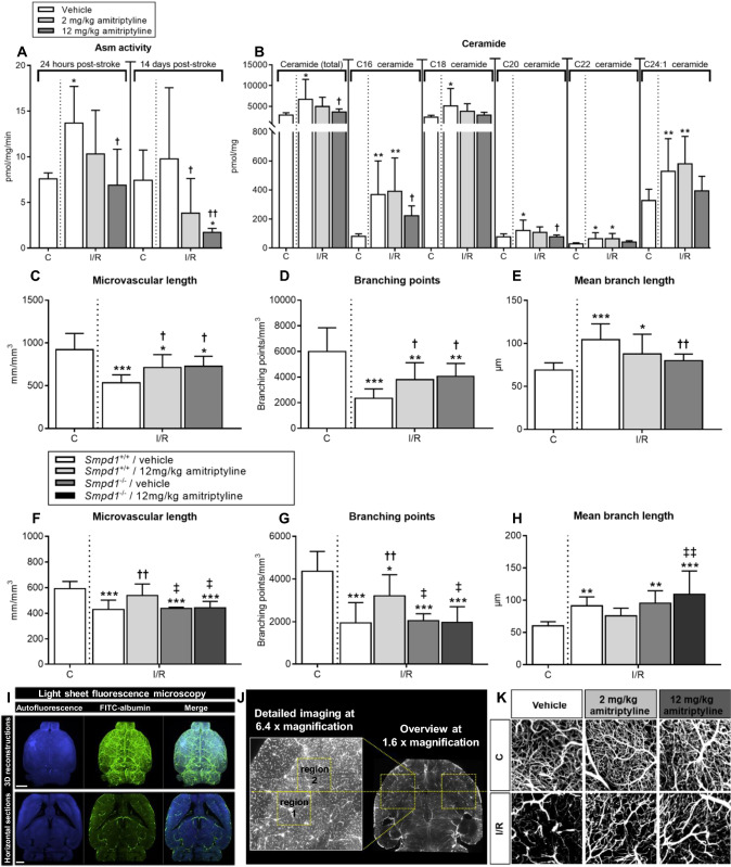
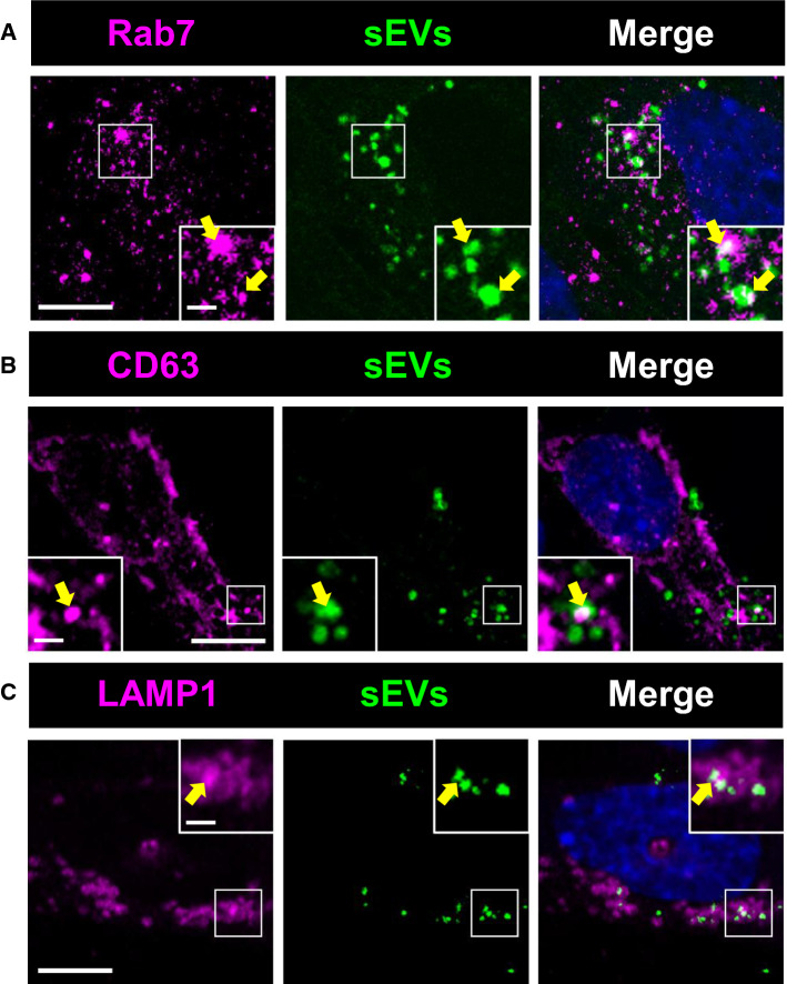
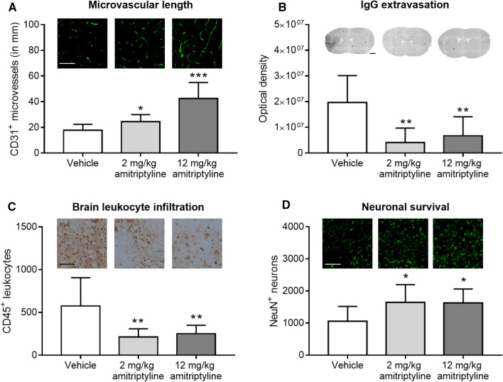
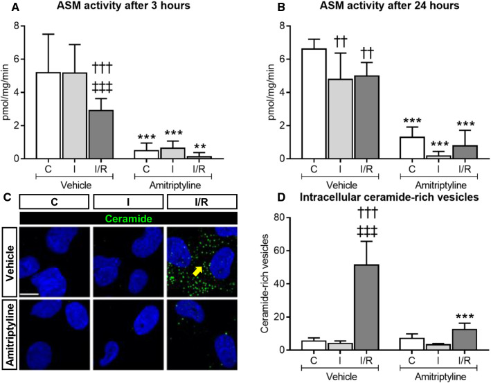
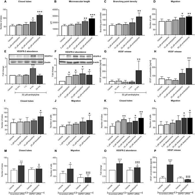
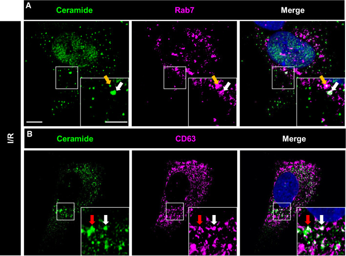
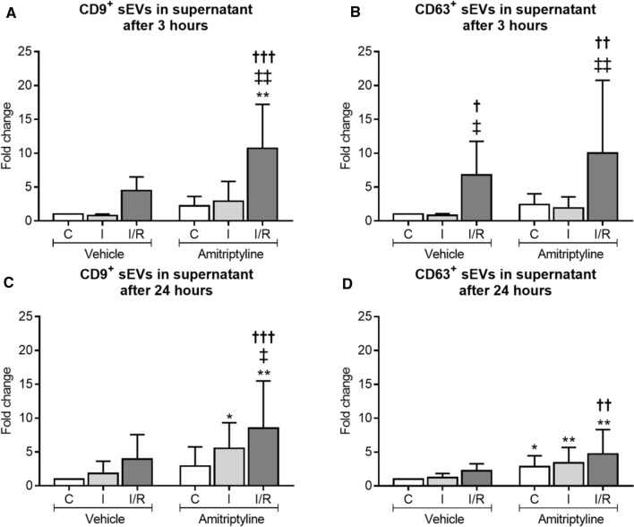
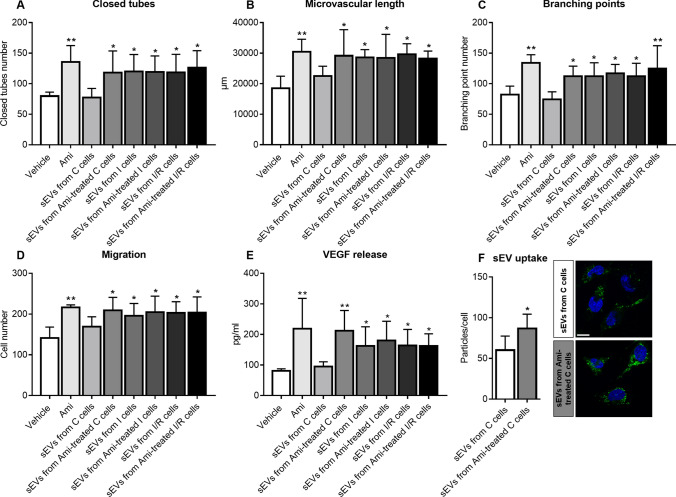
Antidepressants have been reported to enhance stroke recovery independent of the presence of depressive symptoms. They have recently been proposed to exert their mood-stabilizing actions by inhibition of acid sphingomyelinase (ASM), which catalyzes the hydrolysis of sphingomyelin to ceramide. Their restorative action post-ischemia/reperfusion (I/R) still had to be defined. Mice subjected to middle cerebral artery occlusion or cerebral microvascular endothelial cells exposed to oxygen-glucose deprivation were treated with vehicle or with the chemically and pharmacologically distinct antidepressants amitriptyline, fluoxetine or desipramine. Brain ASM activity significantly increased post-I/R, in line with elevated ceramide levels in microvessels. ASM inhibition by amitriptyline reduced ceramide levels, and increased microvascular length and branching point density in wildtype, but not sphingomyelinase phosphodiesterase-1 ([Smpd1]) (i.e., ASM-deficient) mice, as assessed by 3D light sheet microscopy. In cell culture, amitriptyline, fluoxetine, and desipramine increased endothelial tube formation, migration, VEGFR2 abundance and VEGF release. This effect was abolished by Smpd1 knockdown. Mechanistically, the promotion of angiogenesis by ASM inhibitors was mediated by small extracellular vesicles (sEVs) released from endothelial cells, which exhibited enhanced uptake in target cells. Proteomic analysis of sEVs revealed that ASM deactivation differentially regulated proteins implicated in protein export, focal adhesion, and extracellular matrix interaction. In vivo, the increased angiogenesis was accompanied by a profound brain remodeling response with increased blood-brain barrier integrity, reduced leukocyte infiltrates and increased neuronal survival. Antidepressive drugs potently boost angiogenesis in an ASM-dependent way. The release of sEVs by ASM inhibitors disclosed an elegant target, via which brain remodeling post-I/R can be amplified.
抗抑郁药被报道可独立于抑郁症状存在而增强中风恢复。最近有人提出,它们通过抑制酸性鞘磷脂酶(ASM)发挥稳定情绪的作用,ASM 催化鞘磷脂水解为神经酰胺。它们在缺血/再灌注(I/R)后的恢复作用仍有待确定。用载体或用化学和药理学上不同的抗抑郁药阿米替林、氟西汀或去甲替林处理大脑中动脉闭塞或脑微血管内皮细胞缺氧/葡萄糖剥夺的小鼠。I/R 后大脑 ASM 活性显著增加,与微血管中神经酰胺水平升高一致。阿米替林通过抑制 ASM 降低了神经酰胺水平,并增加了野生型小鼠(而非鞘磷脂酶磷酸二酯酶-1([Smpd1])(即 ASM 缺陷型)小鼠的微血管长度和分支点密度,通过 3D 光片显微镜评估。在细胞培养中,阿米替林、氟西汀和去甲替林增加了内皮细胞的管状形成、迁移、VEGFR2 丰度和 VEGF 释放。Smpd1 敲低消除了这种作用。从机制上讲,ASM 抑制剂促进血管生成是通过内皮细胞释放的小细胞外囊泡(sEVs)介导的,这些囊泡在靶细胞中表现出增强的摄取。sEVs 的蛋白质组学分析表明,ASM 失活差异调节了涉及蛋白质输出、焦点粘附和细胞外基质相互作用的蛋白质。在体内,增加的血管生成伴随着深刻的大脑重塑反应,表现为血脑屏障完整性增加、白细胞浸润减少和神经元存活增加。抗抑郁药以 ASM 依赖的方式强力促进血管生成。ASM 抑制剂释放的 sEVs 通过一个优雅的靶点揭示了一种方式,通过这种方式可以放大 I/R 后的大脑重塑。